Full access
Regular Article
Published Online: 1 February 2006

Effects of Childhood Sexual Abuse on Neuropsychological and Cognitive Function in College Women

Publication: The Journal of Neuropsychiatry and Clinical Neurosciences

Abstract

Twenty-six college women with a history of repeated childhood sexual abuse were recruited from the community and compared with 19 healthy female collegiate subjects on neurocognitive measures. Abused subjects showed increased response latency variability and diminished inhibitory capacity during a GO/NO-GO/STOP vigilance task. A strong association was found between duration of abuse and memory impairments. Math Scholastic Aptitude Test (SAT) scores were significantly lower in abused subjects when matched against comparison subjects and when compared to their own Verbal SAT scores. Childhood sexual abuse appears to be associated with a constellation of neuropsychological deficiencies even in a group of relatively healthy women.
The neuropsychological effects of childhood sexual abuse have recently received increased attention. Deficits in short-term verbal memory have been observed in adults with childhood sexual abuse.1 Deficient inhibitory capacity,2 problems with verbally mediated higher cognitive abilities,3 and distractibility and impaired sustained attention4 have been observed in abused children.
Our laboratory, and others, have shown that a history of abuse is related to 1) increased electrophysiological abnormalities, predominantly in the left frontotemporal region5; 2) a higher prevalence of ictal temporal lobe epilepsy-like symptoms6; 3) abnormally elevated left hemisphere EEG coherence7; 4) reduced volume of the left hippocampus811; 5) smaller size of the corpus callosum1215; 6) alterations in the paramagnetic properties of the cerebellar vermis16; 7) abnormalities in cortical size, symmetry, and neuronal density1720; and 8) highly lateralized hemispheric responses to memory recall.21 Together, these findings, and others, support the hypothesis that abuse has a deleterious effect on brain development.
A major limitation of previous studies has been a reliance on clinical samples, which runs the risk of overemphasizing the consequences of childhood sexual abuse. Published imaging and electrophysiological studies have focused on abused subjects with posttraumatic stress disorder (PTSD).7,8,10,12,13 However, only about one-third of individuals with childhood sexual abuse have a lifetime diagnosis of PTSD.22 Hence, past findings may not be applicable to most victims of childhood sexual abuse who never develop PTSD.
Second, a serious unforeseen problem arises as a consequence of restricting trauma studies to subjects with PTSD. Gilbertson et al.23 found that nontrauma exposed monozygotic twins of subjects with combat-related PTSD had reduced hippocampal volume, which strongly suggests that reduced hippocampal volume may be a risk factor for the development of chronic PTSD, rather than a consequence of trauma exposure and PTSD. Hence, studies that only evaluate abused subjects with PTSD cannot disentangle whether observed differences are a consequence of the traumatic stress, or a preexisting risk factor for PTSD. However, if all exposed subjects are examined regardless of diagnosis and outcome, then this conundrum is avoided.
We investigated the effects of childhood sexual abuse on neuropsychiatric status and cognitive development in a nonclinically referred, community-based sample. Young adults, ages 18 to 22 years, were recruited to focus on a circumscribed period of brain maturation. Based on previous literature, we hypothesized that young adults with childhood sexual abuse would have problems with memory recall,1,24 left hemisphere processing,5 left-right hemispheric integration,21 and inhibitory capacity.25,26

METHOD

Participants

Subjects were recruited via advertisements (i.e., bulletin board postings, newspaper ads, and subway and bus ads) looking for healthy right-handed individuals ages 18 to 22 interested in participating in “psychiatric research.” Subjects (N=732) were initially screened with 1) telephone interviews to obtain basic demographic information and ascertain whether any exclusion criteria were met; 2) rating scales to assess current psychiatric symptoms and parent–child attachment; and 3) questionnaires to ascertain family history of psychopathology and lifetime history of exposure to traumatic stressors. The primary entry criterion was a history of three or more episodes of “forced contact sexual abuse before their 18th year and at least 2 years prior to enrollment. An abusive episode was defined as one in which the subject was forced against her will into contact with the sexual part of her body or the perpetrator’s body.” The contact had to be accompanied by threats of harm to self or others, or feelings of fear or terror. Details of the abuse were ascertained through the use of the Traumatic Antecedents Questionnaire.27
Other criteria for participating included: 1) absence of any alcohol, drug, or medication use for at least 2 weeks; 2) excellent hearing; and 3) good medical health. Potential subjects were excluded if they presented with a history of medical disorders (including neurological disease/insult, head injury, migraine headaches, and seizures); psychotic disorders; pregnancy; past or present alcohol/substance abuse; premature birth; complications during mother’s pregnancy or delivery; in utero exposure to alcohol or drugs; a history of physical abuse (defined as any degree of intentional injury above the shoulders, or any intentional injury below the shoulders that received or should have received medical attention); or exposure to any other forms of trauma (e.g., motor vehicle accidents, natural disasters, near drowning, witnessing abuse, animal attacks, gang violence).
These criteria were used to obtain a select sample of individuals with childhood sexual abuse who were free of extraneous factors that could adversely affect neurocognitive development. These criteria excluded 95% of the 732 initial respondents. Twenty-eight percent of the subjects who completed all of the prescreening instruments (N=564) had a self-reported history of childhood sexual abuse, but only 9.5% of the prescreened sample had a history of childhood sexual abuse unaccompanied by exposure to other forms of abuse.
The final experimental sample consisted of 29 subjects. Because only three men were in the abused sample, we chose to focus on the 26 abused women (mean age = 20.0 years). Nineteen women with no current or past DSM–IV axis I disorder (based on the Structured Clinical Interview for DSM–IV Axis I Disorders [SCID-I])28 and with no history of abuse nor exposure to other traumatic events served as a very healthy contrast group (mean age = 19.4 years). Subjects were paid for their participation, and the study was approved and monitored by the McLean Hospital Institutional Review Board.

Design and Procedure

Eligible subjects were invited to participate in a neuropsychiatric evaluation. First, subjects were interviewed with the SCID-I. They were then administered the Memory Assessment Scale (MAS)29 a dichotic listening task developed by Springer et al.30 and a computerized continuous performance task developed by the senior author (MHT). The MAS assesses short-term, verbal, and visual memory. Validity studies indicated that MAS scores are sensitive to left versus right hemisphere dysfunction. Patients with left-hemisphere lesions had deficient verbal memory and those with right hemisphere injury had deficient visual memory on this test.29 The Springer dichotic listening task consisted of precisely timed paired words presented to the right and left ears. The words were of equal duration and similar sound (e.g., “boat” and “goat”). This test has been used to assess one component of hemispheric integration. Springer et al.30 found that traumatic brain-injured patients with symptoms suggestive of temporal lobe epilepsy had a diminished capacity to recognize both words. Carbamazepine was found to enhance their capacity to recognize both of the paired words.
The continuous performance task consisted of two parts. The first component was a 5-minute, two-choice GO/NO-GO test designed simply to determine reaction time. Targets and nontargets (50:50) were 8-pointed and 5-pointed stars presented for 500 msec at random screen positions with a fixed 2-sec interstimulus interval. The second component was a GO/NO-GO/STOP continuous performance task. The stop signal was presented as a visual change in the 8-pointed star in which a large red octagon (“stop sign”) appeared in the center of the target between 30 and 330 msec after its appearance. This component was 15 minutes in duration, with a 2-sec interstimulus interval and 500 msec stimulus duration. The stimuli were equally divided between targets, nontargets, and variably delayed stop signals.
Lastly, subjects provided via self-report Scholastic Aptitude Test (SAT) scores, which reflect their verbal and mathematical reasoning skills.

Statistics

To ascertain differences between the abused and healthy groups on neurocognitive functioning, analysis of covariance (ANCOVA) was conducted on the scores from the various measures. Covariate analysis was performed to control for potential moderating effects, such as current symptoms of depression (Hamilton Depression Rating Scale),31 anxiety (Hamilton Anxiety Rating Scale)32 and PTSD (Mississippi PTSD Scale Civilian Version)33,34 and socioeconomic status (SES).35 Linear regression analyses were also performed to ascertain the relationship(s) between the nature/extent of childhood sexual abuse and neurocognitive function.

RESULTS

Abuse history.

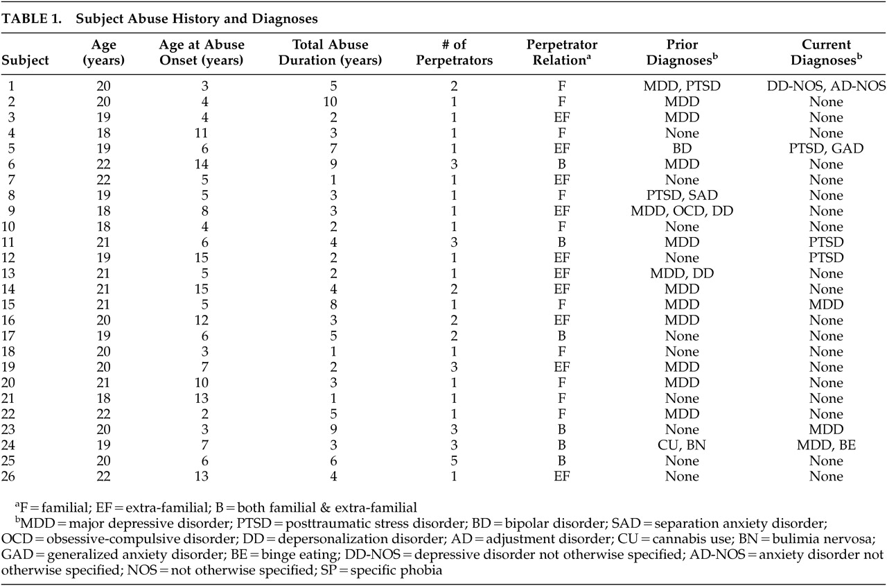
Subjects reported an average of eight sexual abuse incidents (range = 3 to 50+ episodes) with 15 subjects abused by only one perpetrator and the remaining subjects abused by two to five perpetrators (Table 1). A total of 45 perpetrators were reported across subjects. Mean age at abuse onset was 6.3 years (SD = 3.2, range=2–15), and the mean duration of abuse was 3.2 years (SD = 2.2, range=1–10). Subjects in the abuse group had an average Hollingshead SES score of 3.8 (SD = 0.9) versus 4.2 (SD = 0.7) for healthy comparison subjects (F=1.89, df=1, 43, p=0.18)

Diagnostic interview.

Eighteen abused subjects (69%) had a lifetime history of a DSM–IV axis I disorder (Table 1), and seven subjects (27%) had a current axis I disorder. Three subjects (12%) currently met criteria for a major mood disorder, and three subjects met criteria for PTSD (12%). Although all subjects denied a history of substance use on a paper-and-pencil measure, one subject was found on interview to have had a past history of substantial cannabis use during 1 year of high school. No subject had a current substance use problem. Breathalyzer tests and urine drug screens assured that no subject was using drugs at the time of testing.

Neuropsychological Data

Memory testing.

Table 2 displays group scores on the MAS. The abused and comparison groups did not differ in short-term or verbal memory. Intriguingly, childhood sexual abuse subjects exhibited a trend for higher overall visual and global memory scores than healthy comparison subjects. The duration of childhood sexual abuse by the most closely related perpetrator (parent>sibling>uncle>otherfamily>friend>authority figure>stranger/acquaintance) was strongly associated with impairments in all memory categories. Linear regression analyses indicate that for each year of abuse, there was a 2.4 (SD = 0.8%), 2.0 (SD = 1.0%), 1.9 (SD = 0.6%), and 2.3 (SD = 0.5%) reduction in short-term, verbal, visual and global memory scores, respectively (see Table 2 for statistics). Age at abuse onset, the number of perpetrators, and number of years abused by other individuals did not significantly enhance the association between maltreatment and memory performance beyond that accounted for by duration of abuse by the most closely related perpetrator. Symptoms of depression, anxiety, or PTSD or history of these disorders did not influence the strong relationship between duration of abuse by the most closely related perpetrator and measures of memory function.
To ascertain whether specific differences in patterns of memory performance were present, MAS subscale scores were analyzed with ANCOVA using global memory as a covariate to adjust for differences in overall performance. Abused subjects had lower visual span (abuse group: 9.41 [SD = 2.65]; comparison group: 11.96 [SD = 2.71]; F=8.78, df=1, 38, p<0.005) but superior verbal span (abuse group: 12.42 [SD=2.71]; comparison group: 10.59 [SD=2.76], F=4.35, df=1,38, p=0.04). No other significant differences emerged in their pattern of memory performance on the 12 subscales.

Dichotic listening task.

No significant differences emerged between groups on the dichotic listening task for each ear in isolation as well as both in unison. Subjects with childhood sexual abuse correctly identified 41.2 (SD=6.0) of the words presented bilaterally; healthy subjects identified 41.5 [SD=6.0] (F=0.024, df=1,37, p=0.80).

Attention tasks.

On the brief GO/NO-GO task, subjects with childhood sexual abuse performed as well as the healthy group. On the GO/NO-GO/STOP task, abused subjects were more variable in their reaction times (115 msec versus 98 msec, respectively; F=4.56, df=1,32, p=0.04). However, their accuracy and latency scores were not significantly different than healthy subjects (p>0.20). Abused subjects and healthy subjects slowed their reaction time by the same degree (48.1 [SD = 18.8%] and 40.0 [SD = 18.7%]; F=1.53, df=1, 32, p>0.20) to contend with the stop signal.
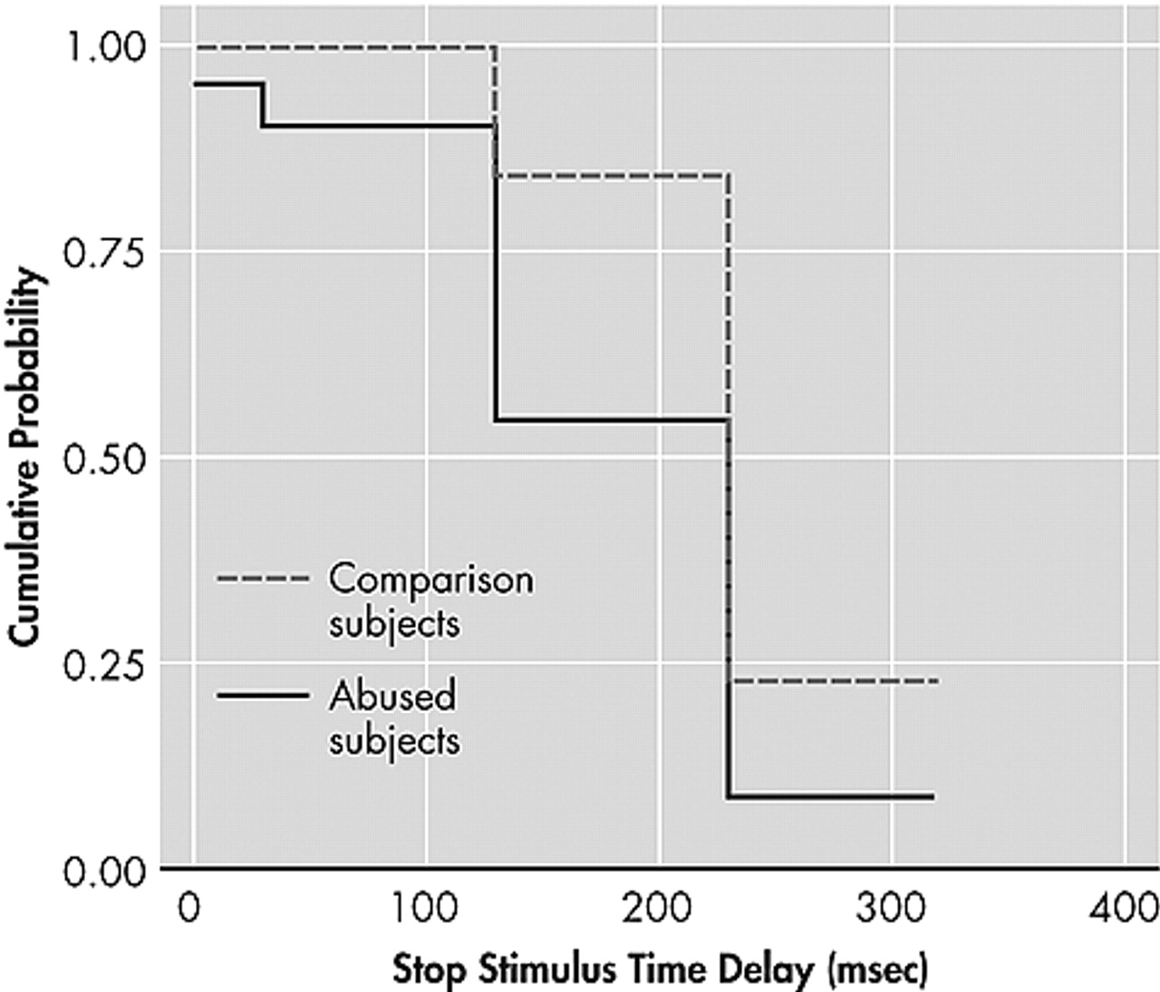
The primary purpose of the STOP task was to assess the capacity to inhibit responses. A survival analysis was conducted on subjects’ failure to sustain near error-free (>95% accuracy) responding as the delay between target presentation and stop signal increased from 30 to 130, 230, and 330 msec. Figure 1 illustrates the calculated survival curve.36
Between-group differences at each interval were ascertained using z tests. At the 30 msec and 130 msec delay intervals, no differences were observed between groups in inhibitory responses (z = −1.05, p>0.2; z = −1.53, p>0.1; respectively). However, a significantly greater percentage of comparison subjects continued to perform nearly flawlessly at the 230 msec (z = −2.2, p = 0.03) and 330 msec delay intervals (z = −2.0, p=0.04).

Scholastic Aptitude Test (SAT).

Self-reported SAT scores were available for 85% of abused subjects and 89% of comparison subjects (two subjects took alternative tests, three could not recall their scores). No significant differences were found between SAT reporters and nonreporters on all of the major MAS subscale scores (all p>0.05). Relative to the healthy group, abused subjects scored 66 points lower on the test (verbal + math, 1221 [SD=26] versus 1287 [SD=31]; F=2.47, df=1, 34, p=0.13). Interestingly, the abused group did not differ from the healthy group on the verbal test (636 [SD=18] versus 628 [SD = 21]; F=0.07, df=1, 34, p>0.60), but scored on average 74 points lower on the math section (585 [SD=14] versus 659 [SD=17], respectively; F=9.3, df=1, 34, p=0.004) after covarying for depression, anxiety, PTSD-associated symptoms, and socioeconomic status. Math SAT scores were strongly affected by a history of childhood sexual abuse, but were not affected by history of depression (F=0.414, df=1, 35, p>0.5) or PTSD (F=0.321, df=1, 35, p>0.5). Within group analyses indicated that the verbal-math difference was significant for abused subjects (t=3.28, df=22, p=0.003) but not for comparison subjects (t=1.29, df=16, p>0.20). A significant correlation was also found between verbal and math SAT scores for abused subjects (r=0.51, p<0.02) but not for comparison subjects (r=0.06, p>0.80).

DISCUSSION

Differences emerged between healthy female college students with no history of trauma or psychiatric illness and a community sample of young adult college women with childhood sexual abuse in inhibitory capacity, memory functions, and scholastic aptitude. A number of factors must be considered when interpreting these findings and comparing them with previous reports. First, we selected subjects with childhood sexual abuse without regard to psychiatric symptoms. Although childhood sexual abuse is a substantial risk factor for later psychopathology,37,38 many children exposed to such abuse never develop any psychiatric condition in general nor PTSD in particular.39,40 Recruitment of abused subjects without regard to psychiatric illness avoids the possibility (discussed in the introduction) that observed alterations were not a consequence of the maltreatment but a risk factor for PTSD or other prespecified diagnosis.23
A second, important aspect of this study was that all of the recruited subjects were college students. Further, they were unmedicated, had no significant history of alcohol use, and had no other exposure to traumatizing events. Studies of older adults are often complicated by varying degrees of drug or alcohol use and by a lifetime history of exposure to other traumatic experiences. Subjects in this study also had a history of repeated, forced, and unwanted sexual contact, but penetration was not required for enrollment. Forced penetration may be an important factor related to the degree of adverse psychiatric outcome.41,42
Overall, these selection criteria biased the sample to include a substantial number of less traumatized subjects with good psychiatric health and above-average cognitive capacity. Subjects were intentionally recruited in this manner to provide a rigorous challenge to the hypothesis that childhood sexual abuse is accompanied by neuropsychiatric and neurocognitive abnormalities. Hence, observed differences between the abused sample and healthy comparison subjects are particularly compelling.
These findings are in accord with some previous reports. First, diminished inhibitory capacity was reported by Mezzacappa et al.2 using a GO/NO-GO/STOP continuous performance task. Their sample consisted of abused boys who were enrolled in therapeutic schools for children with emotional and behavioral problems. The present study suggests that alterations in inhibitory capacity may transcend age, sex, and educational setting.
Abused subjects in the present study had deficits in inhibitory capacity, but no evidence emerged of impaired sustained attention or distractibility on the continuous performance task. Beers and De Bellis4 found that abused children performed more poorly on measures of freedom from distractibility and sustained visual attention. However, they focused specifically on abused children with PTSD and used different means to study attention.
Various cognitive deficits have been reported in several studies, including short-term memory deficits,1,43 and lower levels of intellectual ability, academic attainment, abstract reasoning, and executive function.4,44,45 Koenen et al.46 found that children exposed to high levels of domestic violence had intelligence scores that were lower than unexposed children and that domestic violence was associated with a dose-dependent intellectual decline independent of latent genetic influences. Similarly, we found a strong graded association between duration of childhood sexual abuse and memory function. No overall difference between abused subjects and the contrast group was found because the majority of subjects had a relatively short duration of abuse. Abused subjects with the most profound memory impairments are probably unlikely to matriculate. It should be noted that extrapolating scores for the abuse group back to the zero abuse point (regression intercept) yields mean MAS scores 3.6% to 9.8% greater than comparison subjects (Table 2). If it were true that childhood sexual abuse produces a duration-dependent decrease in memory function, then some of the abused subjects might have had high MAS scores if they had never been abused. This does not imply that individuals with superior memory abilities are at greater risk for abuse, but that by attenuating memory ability abuse may reduce an individuals’ likelihood of attending college, suggesting that those who attend may have had greater than average innate ability.
The selective deficit in math SAT scores in the abused group is interesting, as is the observation that abused subjects had a significant correlation between their verbal and math SAT scores whereas comparison subjects did not. Imaging and lesion studies have provided strong evidence for the involvement of left frontal regions in mathematical abilities.4750 However, evidence also exists for bilateral processing with the two hemispheres contributing different capabilities. For instance, the whole left dorsolateral frontal cortex was activated when a verbal strategy was used to perform subtractions, whereas both right and left prefrontal regions were activated when a visual imagery strategy was utilized.51 Dehaene et al.52 demonstrated that exact arithmetic is left lateralized and recruits networks involved in word-association processing, but approximate arithmetic shows language independence, relies on a sense of numerical magnitudes, and recruits bilateral areas of the parietal lobes involved in visuospatial processing. The math SAT questions focus on algebra, geometry, and magnitude estimation and have been designed so that they can be answered correctly using both exact and approximate approaches.
With these considerations in mind, we hypothesize that the lower math SAT scores of the abused sample could stem from a defect in hemispheric integration. Schiffer et al.21 found that subjects with a history of childhood abuse predominantly activate their left hemisphere while recalling neutral material, and predominantly activate their right hemisphere when recalling disturbing personal events. Hence, subjects with a childhood abuse history may be strongly reliant on left hemisphere function during the SAT, and may be less able to utilize more right hemisphere-based visuospatial mathematical skills that could enhance test performance. The hypothesized reliance of abused subjects on left hemisphere-based mathematical skills involved with word-association is consistent with the observation that math and verbal SAT scores were correlated in the abused group but not in the comparison subjects. Reduced hemispheric integration may be a consequence of reduced corpus callosal area that has been observed in abused children.1215
There are several significant limitations to this study. First, these subjects were less symptomatic and possibly less severely affected by childhood sexual abuse than subjects in other studies. One reason for this is that we limited the abused sample to subjects who had experienced childhood sexual abuse as their only traumatic experience. Exposure to disparate traumatic events is a common occurrence, and it appears to exert an additive or even synergistically deleterious effect.6 Limiting the sample in this way, however, provides strong evidence that identified group differences are related to childhood sexual abuse and not to another type of maltreatment, nor to the interaction of exposure to multiple traumas.
Second, there is the relatively narrow focus of the instruments utilized. We intentionally limited testing to instruments for which we had specific predictions and chose not to include a comprehensive test battery to avoid the statistical problems that accompany global explorations. The results of the present study show that neurocognitive differences exist that are consistent with observations made using imaging and electrophysiological techniques. They do not, however, detail the full breadth of possible neuropsychological differences.
The third limitation is the use of self-report for SAT scores. Subjects were not required to provide official documentation as proof of their SAT performance. However, the potential validity of self-report SAT scores is supported in the literature. For example, in a sample of predominantly white women, Cassady53 found quite high correlations between self-report and actual performance for the SAT total score (r = 0.88, p=0.0001), verbal scale (r = 0.73, p=0.0001), and math scale (r = 0.89, p=0.0001). Nonetheless, our preliminary finding of greatly diminished SAT math scores of the abused group needs to be replicated in a separate sample by directly evaluating mathematical abilities with a standardized achievement test.
The fourth limitation is the exclusive focus on young women. Several studies suggest that early maltreatment may exert an even more deleterious effect on men.13,15 Further research is needed to ascertain whether boys exposed to childhood sexual abuse show the same constellation of neuropsychological differences in adulthood. It must also be noted that associations between childhood sexual abuse and altered neurocognitive function are correlational. We cannot exclude the possibility that these subjects had preexisting abnormalities that increased their risk of being sexually abused, although it is difficult to imagine very young children bringing forth their own sexual abuse.
The fifth limitation is that the onset of abuse occurred during widely different stages of neural development. We have speculated that a given brain region is most vulnerable to insult (e.g., traumatic stress) during phases of most rapid development.54,55 Accordingly, we have preliminary data suggesting that childhood sexual abuse during early childhood has a negative impact on gray matter volume of more primitive brain regions whereas later childhood sexual abuse (e.g., adolescence) affects primarily neocortical areas. However, many brain regions, including the corpus callosum,56 hippocampus,57 and frontal regions,58 gradually myelinate over decades and this process appears vulnerable to the effects of stress. Myelination depends on glial cell division,59 which occurs throughout life, and stress hormones suppress the critical final mitosis of granule cells from proliferative precursors into postmitotic Schwann cells and oligodendrocytes60 that form the myelin sheath. Hence, duration of abuse may be a more critical factor influencing white matter development than age of abuse, and can provide an explanation for why abuse at different ages might exert similar effects on certain neurocognitive measures.
Last, we speculate that the observed math deficit and relative deficiency in visuospatial span in childhood sexual abuse subjects is analogous to individuals with nonverbal learning disabilities, specifically arithmetic learning disabilities.61 That is, both are presumed to have problems with right-hemispheric processing. We further speculate, however, that the apparent right hemisphere deficiency in childhood sexual abuse subjects is more directly related to a disruption in right-left hemisphere integration,61 which may be a consequence of early stress effects on the developing corpus callosum.15,19,62 If this were true, then it might follow that these individuals would benefit from efforts to foster right-left hemisphere integration. We have postulated that this might be possible through training in tasks that normally require precise right-left hemisphere interactions, such as learning a musical instrument.63 Schiffer64 has been working on an alternative approach using lateral visual field stimulation to preferentially activate right versus left hemispheric cortical regions,65 which he has found useful in fostering integration in the course of psychotherapy. Overall, this study provides additional evidence that childhood sexual abuse is associated with neurocognitive abnormalities, and that these abnormalities are discernible in a healthy collegiate sample, are not a consequence of differences in socioeconomic status, or attributable to residual symptoms of depression, anxiety, or PTSD.
ACKNOWLEDGMENTS

Funding for this study was provided by RO1 awards MH-53636 and MH-66222 from the NIMH, and DA-016934 and DA-017846 from the National Institute on Drug Abuse to MHT.
FIGURE 1.  Survival Analysis Indicating Fraction of Abused and Comparison Subjects Who Continue to Perform Almost Flawlessly by Inhibiting Response to a CPT Target That Turns Into a Stop Target After Variable Delay
TABLE 1. Subject Abuse History and Diagnosis
TABLE 2. Memory Assessment Scale Scores and Regression Analysis of Abuse Duration Effects

References

1.
Bremner JD, Randall P, Scott TM, et al: Deficits in short-term memory in adult survivors of childhood abuse. Psychiatry Res 1995; 59(1-2):97-107
2.
Mezzacappa E, Kindlon D, Earls F: Child abuse and performance task assessments of executive functions in boys. J Child Psychol & Psychiatry & Allied Disciplines 2001; 42:1041–1048
3.
Palmer LK, Frantz CE, Armsworth MW, et al: Neuropsychological sequelae of chronically psychologically traumatized children: Specific findings in memory and higher cognitive functions, in Trauma & Memory. Edited by Williams LM. Thousand Oaks, Calif, Sage Publications, 1999, pp 229-244
4.
Beers SR, De Bellis MD: Neuropsychological function in children with maltreatment-related posttraumatic stress disorder. Am J Psychiatry 2002; 159:483–486
5.
Ito Y, Teicher MH, Glod CA, et al: Increased prevalence of electrophysiological abnormalities in children with psychological, physical, and sexual abuse. J Neuropsychiatry Clin Neurosci 1993; 5:401–408
6.
Teicher MH, Glod CA, Surrey J, et al: Early childhood abuse and limbic system ratings in adult psychiatric outpatients. J Neuropsychiatry Clin Neurosci 1993; 5:301–306
7.
Ito Y, Teicher MH, Glod CA, et al: Preliminary evidence for aberrant cortical development in abused children: a quantitative EEG study. J Neuropsychiatry Clin Neurosci 1998; 10:298–307
8.
Bremner JD, Randall P, Vermetten E, et al: Magnetic resonance imaging-based measurement of hippocampal volume in posttraumatic stress disorder related to childhood physical and sexual abuse-a preliminary report. Biol Psychiatry 1997; 41:23–32
9.
Driessen M, Herrmann J, Stahl K, et al: Magnetic resonance imaging volumes of the hippocampus and the amygdala in women with borderline personality disorder and early traumatization. Arch Gen Psychiatry 2000; 57:1115–1122
10.
Stein MB, Koverola C, Hanna C, et al: Hippocampal volume in women victimized by childhood sexual abuse. Psychol Med 1997; 27:951–959
11.
Vythilingam M, Heim C, Newport J, et al: Childhood trauma associated with smaller hippocampal volume in women with major depression. Am J Psychiatry 2002; 159:2072–2080
12.
Teicher MH, Ito Y, Glod CA, et al: Preliminary evidence for abnormal cortical development in physically and sexually abused children using EEG coherence and MRI. Ann N Y Acad Sci 1997; 821:160–175
13.
De Bellis MD, Keshavan MS, Clark DB, et al: A.E. Bennett Research Award. Developmental traumatology, part II: brain development. Biol Psychiatry 1999; 45:1271–1284
14.
De Bellis MD, Keshavan MS, Shifflett H, et al: Brain structures in pediatric maltreatment-related posttraumatic stress disorder: a sociodemographically matched study. Biol Psychiatry 2002; 52:1066–1078
15.
Teicher MH, Dumont NL, Ito Y, et al: Childhood neglect is associated with reduced corpus callosum area. Biol Psychiatry 2004; 56:80–85
16.
Anderson CM, Teicher MH, Polcari A, et al: Abnormal T2 relaxation time in the cerebellar vermis of adults sexually abused in childhood: potential role of the vermis in stress-enhanced risk for drug abuse. Psychoneuroendocrinology 2002; 27(1-2):231-244
17.
Carrion VG, Weems CF, Eliez S, et al: Attenuation of frontal asymmetry in pediatric posttraumatic stress disorder. Biol Psychiatry 2001; 50:943–951
18.
De Bellis MD, Keshavan MS, Frustaci K, et al: Superior temporal gyrus volumes in maltreated children and adolescents with PTSD. Biol Psychiatry 2002; 51:544–552
19.
De Bellis MD, Keshavan MS, Shifflett H, et al: Brain structures in pediatric maltreatment-related posttraumatic stress disorder: a sociodemographically matched study. Biol Psychiatry 2002; 52:1066–1078
20.
De Bellis MD, Keshavan MS, Spencer S, et al: N-acetylaspartate concentration in the anterior cingulate of maltreated children and adolescents with PTSD. Am J Psychiatry 2000; 157:1175–1117
21.
Schiffer F, Teicher MH, Papanicolaou AC: Evoked potential evidence for right brain activity during the recall of traumatic memories. J Neuropsychiatry Clin Neurosci 1995; 7:169–175
22.
Widom CS: Posttraumatic stress disorder in abused and neglected children grown up. Am J Psychiatry 1999; 156:1223–1229
23.
Gilbertson M, Shenton M, Ciszewski A, et al: Smaller hippocampal volume predicts pathologic vulnerability to psychological trauma. Nat Neurosci 2002; 5:1242–1247
24.
Bremner JD, Randall P, Vermetten E, et al: Magnetic resonance imaging-based measurement of hippocampal volume in posttraumatic stress disorder related to childhood physical and sexual abuse-a preliminary report. Biol Psychiatry 1997; 41:23–32
25.
Mezzacappa E, Kindlon D, Earls F: Child abuse and performance task assessments of executive functions in boys. J Child Psychol Psychiatry 2001; 42:1041–1048
26.
Teicher MH, Andersen SL, Polcari A, et al: The neurobiological consequences of early stress and childhood maltreatment. Neuroscience and Biobehavioral Reviews 2003; 27(1-2):33-44
27.
Herman JL, van der Kolk BA: Traumatic Antecedents Questionnaire. Cambridge, Mass, Cambridge Hospital, 1990
28.
First MB, Spitzer RL, Gibbon M, et al: Structured Clinical Interview for DSM-IV Axis I Disorders—Clinical Version (SCID-CV). Washington, DC, American Psychiatric Publishing, Inc 1997
29.
Williams JM: Memory Assessment Scales: Professional Manual. Odessa, Fla, Psychological Assessment Resources, 1991
30.
Springer JA, Garvey MJ, Varney NR, et al: Dichotic listening failure in dysphoric neuropsychiatric patients who endorse multiple seizure-like symptoms. J Nerv Ment Dis 1991; 179:459–467
31.
Hamilton M: A rating scale for depression. J Neurol Neurosurg Psychiatry 1960; 23:56–62
32.
Hamilton M: The assessment of anxiety states by rating. Br J Med Psychol 1959; 32:50–55
33.
Vreven DL, Gudanowski DM, King LA, et al: The civilian version of the Mississippi PTSD scale: a psychometric evaluation. J Trauma Stress 1995; 8:91–109
34.
Norris FH, Perilla JL: The revised civilian Mississippi Scale for PTSD: reliability, validity, and cross-language stability. J Trauma Stress 1996; 9:285–298
35.
Hollingshead AB: Four Factor Index of Social Status. New Haven, Conn., Yale University, 1975
36.
Streiner DL: Stayin’ alive: an introduction to survival analysis. Can J Psychiatry 1995; 40:439–444
37.
Dinwiddie S, Heath AC, Dunne MP, et al: Early sexual abuse and lifetime psychopathology: a co-twin-control study. Psychol Med 2000; 30:41–52
38.
Nelson EC, Heath AC, Madden PA, et al: Association between self-reported childhood sexual abuse and adverse psychosocial outcomes: results from a twin study. Arch Gen Psychiatry 2002; 59:139–145
39.
Paolucci EO, Genuis ML, Violato C: A meta-analysis of the published research on the effects of child sexual abuse. J Psychol 2001; 135:17–36
40.
Putnam FW: Ten-year research update review: child sexual abuse. J Am Acad Child Adolesc Psychiatry 2003; 42:269–278
41.
Bulik CM, Prescott CA, Kendler KS: Features of childhood sexual abuse and the development of psychiatric and substance use disorders. Br J Psychiatry 2001; 179:444–449
42.
Kendall-Tackett KA, Williams LM, Finkelhor D: Impact of sexual abuse on children: a review and synthesis of recent empirical studies. Psychol Bull 1993; 113(1):164–180
43.
Stein MB, Kennedy CM, Twamley EW: Neuropsychological function in female victims of intimate partner violence with and without posttraumatic stress disorder. Biol Psychiatry 2002; 52:1079–1088
44.
Carrey NJ, Butter HJ, Persinger MA, et al: Physiological and cognitive correlates of child abuse. J Am Acad Child Adol Psychiatry 1995; 34:1067–1075
45.
Perez CM, Widom CS: Childhood victimization and long-term intellectual and academic outcomes. Child Abuse Negl 1994; 18:617–633
46.
Koenen KC, Moffitt TE, Caspi A, et al: Domestic violence is associated with environmental suppression of IQ in young children. Dev Psychopathol 2003; 15:297–311
47.
Lucchelli F, De Renzi E: Primary dyscalculia after a medial frontal lesion of the left hemisphere. J Neurol Neurosurg Psychiatry 1993; 56:304–307
48.
Tohgi H, Saitoh K, Takahashi S, et al: Agraphia and acalculia after a left prefrontal (F1, F2) infarction. J Neurol Neurosurg Psychiatry 1995; 58:629–632
49.
Burbaud P, Degreze P, Lafon P, et al: Lateralization of prefrontal activation during internal mental calculation: a functional magnetic resonance imaging study. J Neurophysiol 1995; 74:2194–2200
50.
Delazer M, Domahs F, Bartha L, et al: Learning complex arithmetic-an FMRI study. Brain Res Cogn Brain Res 2003; 18:76–88
51.
Burbaud P, Camus O, Guehl D, et al: Influence of cognitive strategies on the pattern of cortical activation during mental subtraction. A functional imaging study in human subjects. Neurosci Lett 2000; 287:76–80
52.
Dehaene S, Spelke E, Pinel P, et al: Sources of mathematical thinking: behavioral and brain-imaging evidence. Science 1999; 284:970–974
53.
Cassady JC: Self-reported GPA and SAT scores, in ERIC Digest, ERIC Clearinghouse on Assessment and Evaluation, College Park, Md.
54.
Teicher MH, Ito Y, Glod CA, et al: Neurophysiological mechanisms of stress response in children, in Severe Stress and Mental Disturbance in Children. Edited by Pfeffer C. Washington, D.C, American Psychiatric Press, 1996, pp 59-84
55.
Teicher MH, Andersen SL, Polcari A, et al: The neurobiological consequences of early stress and childhood maltreatment. Neurosci Biobehav Rev 2003;27(1-2):33-44
56.
Schaefer GB, Thompson JN, Jr, Bodensteiner JB, et al: Quantitative morphometric analysis of brain growth using magnetic resonance imaging. J Child Neurol 1990; 5:127–130
57.
Benes FM, Turtle M, Khan Y, et al: Myelination of a key relay zone in the hippocampal formation occurs in the human brain during childhood, adolescence, and adulthood. Arch Gen Psychiatry 1994; 51:477–484
58.
Klingberg T, Vaidya CJ, Gabrieli JD, et al: Myelination and organization of the frontal white matter in children: a diffusion tensor MRI study. Neuroreport 1999; 10:2817–2821
59.
Lauder JM: Hormonal and humoral influences on brain development. Psychoneuroendocrinology 1983; 8:121–155
60.
Bohn MC: Granule cell genesis in the hippocampus of rats treated neonatally with hydrocortisone. Neuroscience 1980; 5:2003–2012
61.
Semrud-Clikeman M, Hynd GW: Right hemispheric dysfunction in nonverbal learning disabilities: social, academic, and adaptive functioning in adults and children. Psychol Bull 1990; 107:196–209
62.
De Bellis MD, Keshavan MS, Clark DB, et al: Developmental traumatology. part II: brain development. Biol Psychiatry 1999; 45:1271–1284
63.
Teicher MH: Wounds that time won’t heal: The neurobiology of child abuse. Cerebrum 2000; 4:50–67
64.
Schiffer F: Affect changes observed with right versus left lateral visual field stimulation in psychotherapy patients: possible physiological, psychological and therapeutic implications. Comp Psychiatry 1997:1-8
65.
Schiffer F, Mottaghy FM, Pandey Vimal RL, et al: Lateral visual field stimulation reveals extrastriate cortical activation in the contralateral hemisphere: an FMRI study. Psychiatry Res 2004; 131:1–9

Information & Authors

Information

Published In

Go to The Journal of Neuropsychiatry and Clinical Neurosciences
The Journal of Neuropsychiatry and Clinical Neurosciences
Pages: 45 - 53
PubMed: 16525070

History

Published online: 1 February 2006
Published in print: February 2006

Authors

Details

Carryl P. Navalta, Ph.D.
Received February 13, 2004; revised December 30, 2004; accepted January 18, 2005. From the Developmental Biopsychiatry Research Program, McLean Hospital, Belmont, Mass. Address correspondence to Dr. Navalta, McLean Hospital, 115 Mill St., Belmont, MA 02478; [email protected] (E-mail).
Ann Polcari, R.N., Ph.D.
Received February 13, 2004; revised December 30, 2004; accepted January 18, 2005. From the Developmental Biopsychiatry Research Program, McLean Hospital, Belmont, Mass. Address correspondence to Dr. Navalta, McLean Hospital, 115 Mill St., Belmont, MA 02478; [email protected] (E-mail).
Danielle M. Webster, R.N., M.S.C.S.
Received February 13, 2004; revised December 30, 2004; accepted January 18, 2005. From the Developmental Biopsychiatry Research Program, McLean Hospital, Belmont, Mass. Address correspondence to Dr. Navalta, McLean Hospital, 115 Mill St., Belmont, MA 02478; [email protected] (E-mail).
Ani Boghossian
Received February 13, 2004; revised December 30, 2004; accepted January 18, 2005. From the Developmental Biopsychiatry Research Program, McLean Hospital, Belmont, Mass. Address correspondence to Dr. Navalta, McLean Hospital, 115 Mill St., Belmont, MA 02478; [email protected] (E-mail).
Martin H. Teicher, M.D., Ph.D.
Received February 13, 2004; revised December 30, 2004; accepted January 18, 2005. From the Developmental Biopsychiatry Research Program, McLean Hospital, Belmont, Mass. Address correspondence to Dr. Navalta, McLean Hospital, 115 Mill St., Belmont, MA 02478; [email protected] (E-mail).

Metrics & Citations

Metrics

Citations

Export Citations

If you have the appropriate software installed, you can download article citation data to the citation manager of your choice. Simply select your manager software from the list below and click Download.

For more information or tips please see 'Downloading to a citation manager' in the Help menu.

Format
Citation style
Style
Copy to clipboard

View Options

View options

PDF/EPUB

View PDF/EPUB

Get Access

Login options

Already a subscriber? Access your subscription through your login credentials or your institution for full access to this article.

Personal login Institutional Login Open Athens login
Purchase Options

Purchase this article to access the full text.

PPV Articles - Journal of Neuropsychiatry and Clinical Neurosciences

PPV Articles - Journal of Neuropsychiatry and Clinical Neurosciences

Not a subscriber?

Subscribe Now / Learn More

PsychiatryOnline subscription options offer access to the DSM-5-TR® library, books, journals, CME, and patient resources. This all-in-one virtual library provides psychiatrists and mental health professionals with key resources for diagnosis, treatment, research, and professional development.

Need more help? PsychiatryOnline Customer Service may be reached by emailing [email protected] or by calling 800-368-5777 (in the U.S.) or 703-907-7322 (outside the U.S.).

Media

Figures

Other

Tables

Share

Share

Share article link

Share