Abstract

Research on novel treatments for major depressive disorder focuses quite deeply on glutamate function, and this research would benefit from a brain-imaging technique that precisely quantified glutamate function. Signs of a specific form of glutamate-related dysfunction that could be targeted by novel therapies were found using novel, state-of-the-art techniques to address this issue.

Abstract

Objective:

Emerging evidence suggests that abnormalities in amino acid neurotransmitter function and impaired energy metabolism contribute to the underlying pathophysiology of major depressive disorder. To test whether impairments in energetics and glutamate neurotransmitter cycling are present in major depression, we used carbon-13 magnetic resonance spectroscopy (13C MRS) to measure these fluxes in individuals diagnosed with major depression relative to healthy comparison subjects.

Method:

Proton (1H) MRS and 13C MRS data were collected for 23 medication-free individuals with major depression and 17 healthy subjects. 1H MRS provided total glutamate and GABA concentrations, and 13C MRS, coupled with intravenous infusion of [1-13C]glucose, provided measures of the neuronal tricarboxylic acid cycle for mitochondrial energy production, GABA synthesis, and glutamate/glutamine cycling from voxels situated in the occipital cortex.

Results:

Mitochondrial energy production of glutamatergic neurons was 26% lower in the depression group. Paradoxically, no difference was found in the rate of the glutamate/glutamine cycle (Vcycle). A significant correlation was observed between glutamate concentrations and Vcycle in the overall sample.

Conclusions:

The authors interpret the reduction in mitochondrial energy production as being due to either mitochondrial dysfunction or a reduction in proper neuronal input or synaptic strength. Future MRS studies could help distinguish these possibilities.
Major depressive disorder is a debilitating and, in some cases, potentially life-threatening psychiatric disorder. Major depression affects an estimated 350 million people worldwide and is a leading cause of years lost due to disability (1). However, despite the high prevalence and immense economic burden associated with major depression, relatively little is known about the pathophysiology underlying the disorder. Although the monoaminergic neurotransmitter systems have been the primary focus in the majority of investigations exploring the associated pathophysiology for the past five decades, evidence is rapidly accumulating to suggest that the amino acid neurotransmitter systems and impaired mitochondrial function contribute to pathophysiological mechanisms of mood and other stress-related disorders (2, 3).
Clinical studies using magnetic resonance spectroscopy (MRS) have repeatedly shown abnormal amino acid neurotransmitter (glutamate, glutamine, and GABA) content in the brains of patients diagnosed with mood disorders. A meta-analysis of proton (1H) MRS studies found evidence of both diffuse and region-specific changes in glutamine and glutamate content (4), and the recent discovery of significant antidepressant effects associated with drugs targeting the glutamatergic system has led to speculation on the contributions of the system to the pathophysiology of mood disorders (5). In addition to evidence linking glutamate to mood disorders, there is a long history of studies suggesting that abnormalities in the GABA-ergic system contribute to the pathophysiology of mood disorders (6). Early reports suggested that GABA levels were reduced in the blood and cerebrospinal fluid of patients with depression (7). Imaging studies have provided fairly consistent reports suggesting that GABA content is reduced in the brains of patients with depression, especially in the occipital cortex (8, 9), although this appears to occur primarily in certain subtypes of the disorder. Additional studies have suggested that altered function of the amino acid neurotransmitter systems contributes to changes in cortical excitability and inhibition as well as several other pathophysiological processes related to mood disorders (10, 11).
One potential pathophysiological mechanism that could be related to the findings of altered amino acid neurotransmitter system content and function in individuals with mood disorders is impairment in astrocyte function. Astrocytes provide a primary means of glutamate and GABA clearance through the excitatory amino acid transporters 1 and 2 and GABA transporters 1–3, respectively (12, 13). Astrocytes also play a critical role in the metabolism of both glutamate and GABA. Once glutamate is brought into an astrocyte, it is rapidly converted into glutamine by glutamine synthetase. The glutamine is then transported back to glutamatergic neurons to replenish glutamate stores by the mitochondrial enzyme glutaminase in a process referred to as the glutamate/glutamine cycle (14). Glutamine is also transported to GABA-ergic cells, where it is first converted into glutamate by glutaminase and then to GABA by glutamic acid decarboxylase in the GABA/glutamine cycle. Evidence that astrocytic pathology may be associated with mood disorders, along with rodent models of mood disorders (see references 15, 16 for reviews), suggests that the abnormalities observed in the amino acid neurotransmitter systems associated with mood disorders could be secondary to changes in the glutamate/glutamine and GABA/glutamine cycles.
Mitochondria, in addition to serving a critical role in cellular energy metabolism, are also intimately involved in amino acid metabolism and brain function. Work over the past two decades has shown that there is a linear relationship between changes in the rates of neurotransmitter glutamate release and recycling and the neuronal tricarboxylic acid (TCA) cycle, with approximately 80% of neuronal energy metabolism devoted to this function in the resting awake cerebral cortex (17). Therefore, even relatively minor mitochondrial impairment could have an adverse impact on glutamate neurotransmission and brain function.
Given the rapidly mounting evidence suggesting that altered mitochondrial function and amino acid metabolism are associated with depression and other mood disorders (3), we used carbon-13 MRS (13C MRS) to determine whether the rate of the neuronal TCA cycle (VTCAN), the glutamate/glutamine cycle (Vcycle), and GABA synthesis (VGAD) are altered in major depressive disorder. We found a significant reduction in the rate of the neuronal TCA cycle in glutamatergic neurons, implicating the glutamatergic system and mitochondrial energy metabolism as having an important role in the pathology of major depression. Paradoxically, we also found that Vcycle was similar in depressed and healthy comparison subjects, implying a possible alteration in neuronal coupling. To our knowledge, this is the first study to use 13C MRS in vivo to study the pathophysiology of major depression, and it provides the first measures of VGAD in humans as well as the first correlations between 13C MRS and 1H MRS measures of metabolite concentrations in humans.

Method

Participants

Men and women 18–65 years of age were recruited into the study. An institutional review board at Yale University approved all study procedures. Participants provided written informed consent after receiving a complete description of the study. After comprehensive medical and psychiatric assessments, volunteers meeting the study criteria underwent a 1H MRS scan and then a 13C MRS scan, each on a separate day. Two groups of participants were enrolled: a group with major depressive disorder and a healthy comparison group. Participants with major depression had to be medication free for at least 4 weeks; meet DSM-IV criteria for major depressive disorder, confirmed by structured interview with the patient version of the Structured Clinical Interview for DSM-IV (SCID); and have a score >21 on the 21-item Hamilton Depression Rating Scale (HAM-D). Healthy comparison subjects had to have no personal or first-degree family history of an axis I DSM-IV disorder, confirmed by the non-patient version of the SCID. Comparison subjects were age- and sex-matched to the depression group. The exclusion criteria for both groups included a history of or a current major medical or neurological illness, a history of or current substance abuse or dependence, current use of nicotine, any implanted metal, and a current pregnancy. Rating scales included the HAM-D, the Beck Depression Inventory (BDI), and the Hamilton Anxiety Rating Scale (HAM-A).

1H MRS Acquisition and Processing

1H MRS scanning in the occipital cortex was performed as previously outlined (9). Briefly, metabolite levels were measured in a 13.5 cm3 voxel (3.0×1.5×3.0 cm) situated across the midline of the brain, centered 2 cm from the dura. Cortical GABA content was determined using J-editing, in which subspectra were acquired with 8,000 data points over a 410-ms acquisition, with a 2.5-second repetition time and an echo time of 68 ms on a 4-T Bruker spectrometer at the Yale Magnetic Resonance Research Center, with data averaged in 20-second blocks for 20 minutes. Glutamate and glutamine were measured simultaneously using the unedited subspectra of the J-editing acquisition, with in-house software that uses an LCModel approach (18). The metabolites fitted included GABA, glutamate, glutamine, and creatine. The subspectrum obtained without the editing pulse was fitted simultaneously with the J-edited difference spectrum of GABA. Because of limited resolution in vivo, the results for N-acetylaspartate and N-acetylaspartylglutamate were combined and recorded as N-acetylaspartate; creatine and phosphocreatine were combined and recorded as creatine; and the three choline-containing compounds were combined and recorded as choline. This implementation had no macromolecular contamination of GABA (19), so the basis set for fitting did not include a macromolecular signal. The level of aspartate, although present in the spectra, was poorly determined at the echo time of 68 ms and was not used. Uncertainties of individual measurements were determined using a Monte Carlo analysis (19), in which the least-squares spectral fits were treated with random Gaussian noise whose standard deviation was equal to that of the raw data and refitted, using 20 repetitions to estimate the standard deviations of the uncertainty for each metabolite measure. For each metabolite, a threshold for rejection was set at twice the average noise-based standard deviation of the respective metabolite. GABA levels whose uncertainties were greater than 11%, glutamine levels whose uncertainties were greater than 20%, and glutamate levels whose uncertainties were greater than 16% were not included in subsequent analysis.
To account for potential differences in tissue composition, a series of 3-mm-thick contiguous T1-weighted images were used to quantify gray matter, white matter, and cerebrospinal fluid in the voxel of interest (20). The T1-weighted images were measured using a series of inversion-recovery images that required images of the spatial distribution of the radiofrequency power to overcome the problems associated with radiofrequency inhomogeneity. The mean percentage of solid tissue in the acquired voxel did not differ significantly between groups. Thus, no covariance for tissue content was needed. Similarly, the mean ratio of creatine to water did not differ significantly between groups. Hence, the concentration of brain metabolites was calculated assuming a normal creatine concentration of 9 mmol/kg (21).

13C MRS Acquisition, Processing, and Metabolic Modeling

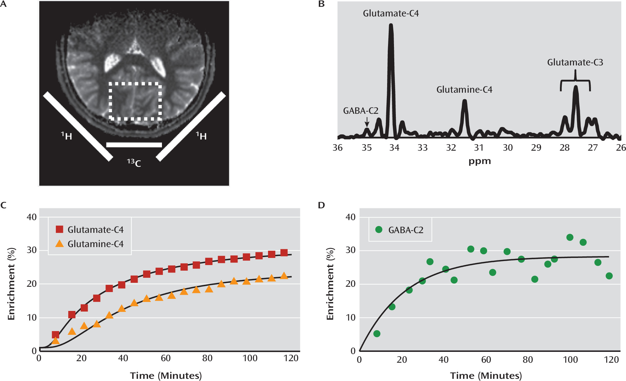
13C MRS acquisition, processing, and kinetic metabolic modeling were performed as described previously (22, 23). In summary, all participants fasted overnight before the 13C MRS acquisition. The 13C MRS studies were performed a mean of 6.7 days (SD=9.91) after the 1H MRS study. In the morning, two intravenous lines were initiated, one for [1-13C]glucose infusion and the other for blood sampling. 13C MRS data were acquired on a 4.0-T magnet. Participants were placed supine, with their heads resting on a radiofrequency probe consisting of one 8.5-cm diameter circular 13C coil and two 12.5-cm diameter, quadrature-driven circular 1H radiofrequency coils. The region of interest was a voxel (5×4×4.5 cm) situated across the midline of the occipital-parietal lobe. After tuning, acquisition of scout images, FASTERMAP shimming, and decoupling power calibration, infusion of [1-13C]glucose was started and 5-minute blocks of 13C MR spectra were acquired for 120 min using polarization transfer (Figure 1). The plasma glucose concentration was rapidly titrated to 180–200 mg/dL and maintained near that level for the duration of 13C MRS acquisition. Plasma glucose concentrations and [1-13C] enrichments were determined from blood samples collected at baseline and during the [1-13C]glucose infusion.
FIGURE 1. Voxel, Spectrum, and Time Courses in Carbon-13 Magnetic Resonance Spectroscopy in a Study of Glutamate Metabolism in Major Depressiona
a Panel A illustrates the placement of the coils and the location of the voxels. Panel B is a graph of the spectrum acquired over 10 minutes at 4 T from the occipital region during [1-13C]glucose infusion at steady state. In panels C and D, the percent enrichment for each metabolite is plotted against time.
Spectral data were analyzed with −2 Hz/6 Hz Lorentzian-to-Gaussian conversion and 16-fold zero-filling followed by Fourier transformation. Software developed in-house with MATLAB, version 7.12.0 (MathWorks, Natick, Mass.) was used to determine the peak heights for the C3 and C4 positions of glutamate, the C4 position of glutamine, and the C2 position of GABA of each spectrum. Peak heights were converted to concentrations of 13C using the fractional enrichment of glutamate C4, determined by isotopomer analysis (24) and the total glutamate concentration measured by 1H MRS. Time courses of glutamate, glutamine, and GABA peaks were analyzed using CWave (25) to implement a three-compartment model of brain metabolism that included astroglia and glutamatergic and GABA-ergic neurons (26). The CWave program iterated the values of the rates of GABA synthesis (VGAD), glutamate neurotransmitter cycling (Vcycle), and the neuronal TCA cycle (VTCAN) to obtain a least-squares fit of the model to the time courses of each participant’s data, using the time courses of the individual’s own plasma glucose concentration and fractional enrichment as input functions. The equations used in the kinetic model are presented in Table S1 in the data supplement that accompanies the online edition of this article.

Statistical Analysis

Prior to each analysis, outcomes were assessed for normality using normal probability plots and Kolmogorov-Smirnov tests. Logarithmic (log10) transformations were performed as necessary on variables with skewed distribution. Independent t tests and chi-square tests were used to determine differences between groups, and Bonferroni correction for multiple comparisons was used where appropriate. Spearman’s rank order was used for correlational analyses. All tests were two-tailed, with the significance threshold set at 0.05.

Results

A total of 46 participants completed all study procedures. Six participants (three in each group) were excluded because of poor 13C MRS spectral quality. Demographic data show that the study participants were well matched for age, sex, and body mass index (BMI) (Table 1). All participants in the depression group were medication free for at least 4 weeks. On average, the clinical characteristics of the depression group are consistent with a moderate level of depression severity with coexisting mild anxiety.
TABLE 1. Demographic and Clinical Characteristics of Participants in a Study of Glutamate Metabolism in Major Depression
CharacteristicMajor Depression Group (N=23)Healthy Comparison Group (N=17)
 MeanSDMeanSD
Age43.010.643.812.8
Body mass index26.34.827.36.6
Hamilton Depression Rating Scale30.15.8  
Beck Depression Inventory26.36.2  
Hamilton Anxiety Rating Scale15.64.8  
 N%N%
Female16701376
First major depressive episode417  
Medication naive835  
Depression subtype    
 Melancholic522  
 Atypical1565  
 None313  

13C MRS Spectra and Metabolic Modeling

In contrast to the more commonly employed 1H MRS methods, 13C MRS is capable of providing unique information about the dynamic processes of metabolism and neurotransmission. Since carbon-12, which is invisible to MRS detection methods, comprises nearly 99% of the carbon content in biological systems, it is possible to track and model the labeling of individual molecules over time with exogenously administered carbon-13, a stable isotope visible by MRS methods. This approach makes it possible to obtain dynamic measures relevant to amino acid neurotransmitter metabolism and neurotransmission. To label the carbon positions of glutamate, glutamine, and GABA, [1-13C]glucose was infused intravenously over 120 minutes during MR spectroscopy acquisition, which was focused on the region of the occipital cortex (Figure 1A). The incorporation of the carbon-13 in glutamate, glutamine, and GABA generates unique signals on the carbon-13 spectrum (Figure 1B). 13C MRS spectra were obtained with a 5-minute time resolution. A plot of the time courses of the carbon-13 labeling in C4 glutamate, glutamine, and C2 GABA is provided in Figures 1C and 1D. In addition, the C3 resonances of glutamate and glutamine were measured and used in the modeling. The steady-state fractional enrichment of glutamine was lower than that of glutamate in most participants, consistent with previous findings (27). Using mass and isotope balance equations, the labeling time courses were used to calculate glutamate/glutamine cycling (Vcycle; a measure of neuronal glutamate release and glial reuptake), neuronal oxidation (VTCAN; mitochondrial energy production specific to glutamatergic neurons), and GABA synthesis (VGAD).

13C MRS Metabolic Fluxes

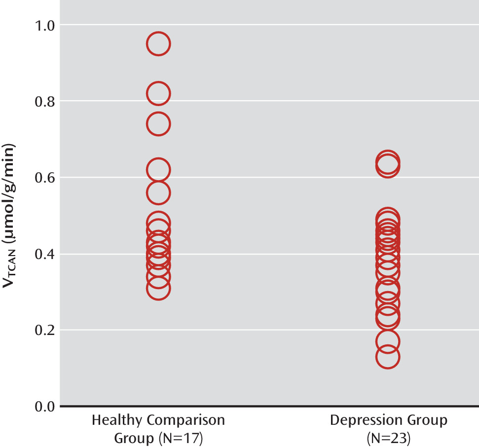
Participants in the depression group had a 26% reduction in oxidative mitochondrial energy production of glutamatergic neurons (depression group: mean VTCAN, 0.35 μmol/g/min [SD=0.14]; healthy comparison group: mean VTCAN, 0.47 μmol/g/min [SD=0.21]; t=2.57, N=40, p=0.01) (Figure 2). Cohen’s d was 0.84 (95% CI=0.80–0.89). The VTCAN differences between groups remained significant after Bonferroni correction for multiple comparisons (p<0.05/3). Vcycle and VGAD did not differ significantly between the two groups (Table 2). The values of Vcycle and VTCAN were consistent with those reported in previous studies (28). The rate of VGAD was approximately 10%–20% of VTCAN, which is consistent with previous animal studies (26, 29).
FIGURE 2. Mitochondrial Energy Production in Participants in a Study of Glutamate Metabolism in Major Depressiona
a VTCAN=neuronal tricarboxylic acid cycle.
TABLE 2. Comparison of Occipital Metabolic Fluxes in a Study of Glutamate Metabolism in Major Depression
 Major Depression Group (N=23)Healthy Comparison Group (N=17)Analysis
MeasureaMeanSDMeanSDtdfp
VTCAN (μmol/g/min)0.350.140.470.212.57380.01b
Vcycle (μmol/g/min)0.190.050.180.04–0.32380.75
VGAD (μmol/g/min)0.040.0140.030.012–0.3534c0.72
a
VTCAN=neuronal tricarboxylic acid cycle; Vcycle=glutamate-glutamine cycle; VGAD=GABA synthesis.
b
Remained significant after adjustment for multiple comparisons.
c
VGAD was excluded for five participants with noise levels higher than 0.05 μmol/g/min.

1H MRS Metabolite Levels

Across all participants, we observed significant correlations between glutamate level and Vcycle (rs=0.45, p=0.004), as well as between glutamate and VTCAN (rs=0.34, p=0.04). However, only the association between glutamate and Vcycle remained significant after Bonferroni correction for multiple comparisons (p<0.05/9). Associations between metabolic fluxes and amino acid neurotransmitter level are listed in Table 3. The mean values of amino acid neurotransmitter level were not significantly different between groups (see Table S2 in the online data supplement). However, we observed an interesting pattern suggesting that GABA levels may have been lower in the subgroup of participants with the melancholic depression subtype (see Figure S1 in the data supplement).
TABLE 3. Correlations Between Metabolic Fluxes and Brain Metabolites in a Study of Glutamate Metabolism in Major Depressiona
MetaboliteVTCANVcycleVGADb
GABA0.230.09–0.06
Glutamine0.160.280.12
Glutamatec0.34*0.45**d0.09
a
Correlations are Spearman’s correlation coefficient. VTCAN=neuronal tricarboxylic acid cycle; Vcycle=glutamate-glutamine cycle; VGAD=GABA synthesis.
b
VGAD was excluded for five participants with noise levels higher than 0.05 μmol/g/min.
c
Two participants had poor spectral fitting for glutamate.
d
Remained significant after correction for multiple comparisons.
*
p<0.05. **p<0.01. p<0.1.

Associations Between Clinical and Spectroscopy Measures in the Depression Group

Number of lifetime major depressive episodes was negatively correlated with glutamate concentration (rs=−0.59, p=0.01). HAM-A scores were negatively correlated with glutamine concentration (rs=−0.47, p=0.03), as well as with VTCAN, although this association fell short of statistical significance (rs=−0.37, p=0.09). However, these correlations did not survive Bonferroni correction for multiple comparisons. Correlations between clinical and spectroscopy measures are detailed in Table S3 in the online data supplement.

Discussion

Using state-of-the-art 13C MRS methods, we studied neuronal oxidative energy production, glutamate-glutamine cycling, and GABA synthesis in individuals with major depression and healthy comparison subjects. We found that oxidative energy production specific to glutamatergic neurons was 26% lower in the depression group. No differences between groups were observed in glutamate-glutamine cycling or GABA synthesis. Total glutamate levels were correlated with glutamate-glutamine cycling and, to a lesser extent, neuronal energy production. However, in contrast to findings in previous studies (8, 9), amino acid neurotransmitters did not differ between groups in this sample. Finally, post hoc exploratory analysis—without correction for multiple comparisons—showed a negative association between glutamate concentration and number of depressive episodes, as well as between glutamine concentration and clinician-rated anxiety scores.
The ability of MRS to separate mitochondrial energy production between glutamatergic neurons, GABA-ergic neurons, and glia is based on the compartmentation of glutamine synthase and glutamic acid decarboxylase in glia and neurons, respectively, and the majority of the neuronal glutamate pool being in glutamatergic neurons (see reference 14 for a review). Because MRS can distinguish carbon-13 labeling in GABA, glutamine, and glutamate, it is possible to use kinetic modeling to obtain separate measures of the TCA cycle in all three compartments (14, 26, 29).
The findings of reduced neuronal energy production in the depression group are generally consistent with reports of slower energy metabolism, primarily decreased baseline levels of β-nucleoside triphosphate (β-NTP) and total NTP (associated with major depression [30, 31]), and with reports from positron emission tomography studies of regional and global reductions in glucose metabolism in major depression (32). In addition, in a rodent model of chronic unpredictable stress, reduced carbon-13 labeling from glucose was observed in glutamate, consistent with findings in human subjects (33).
A potential explanation for the reduced neuronal energy production is mitochondrial impairment in depressed individuals. Mitochondrial dysfunction in mood disorders has been suggested previously (reviewed in reference 3). However, direct in vivo measures of neuronal mitochondrial energy production in major depression have not previously been investigated. Impaired oxidative metabolism, in the absence of reduced neuronal activity, would be consistent with previous evidence suggesting an increase in cerebral glycolysis and lactate, as well as reductions in phosphocreatine level and pH in patients with mood disorders (3, 34).
An alternative explanation is that the reduced neuronal energy production reflects a down-regulation of cortical activity. Extensive animal and human studies have demonstrated a strong coupling, close to 1:1, between energy production in the TCA cycle and the glutamate/glutamine cycle (17). Consistent with this relationship, we observed a positive relationship, approaching statistical significance, between VTCAN and Vcycle (r=0.436, p=0.08) in the healthy comparison subjects in the present study (see Table S4 in the online data supplement). In the depression group, however, the relationship between VTCAN and Vcycle appeared to be weaker (r=0.252, n.s.). This is again reflected in the fact that we observed normal rates of glutamate/glutamine cycling despite slower energy production in the depression group.
The seemingly contradictory findings of reduced neuronal energy metabolism and normal levels of glutamate/glutamine cycling could be reconciled by the presence of an overall reduction of glutamatergic synaptic strength. Brain functional energy needs are largely determined by the ATP needed to maintain ion flows in the post- and presynaptic terminals of excitatory synapses that are coupled to glutamate neurotransmission (17). Reduced overall synaptic strength in individuals with depression would reduce energy demands for the same amount of glutamate-glutamine cycling. In line with this hypothesis, chronic stress has been shown to reduce glutamate AMPA and NMDA receptor expression and transmission, the primary determinants of synaptic strength (35).
Alternatively, the rate of the glutamate/glutamine cycle in the depression group may have been overestimated because of a change in the balance between neuronal and astroglial metabolism in this group. A previous 13C MRS study of healthy aging (22) found that the reduction in neuronal metabolism is accompanied by an increase in glial metabolism, and similar changes in major depression could lead to an overestimate of the rate of the glutamate/glutamine cycle with the kinetic modeling used. Increasing evidence suggests astroglial changes in major depression (16, 36), including in 13C MRS studies in a rodent model of chronic unpredictable stress (33). To overcome this limitation, future studies may employ recently developed carbon-13 methods of combined labeling by 13C-glucose and 13C-acetate (37). Acetate oxidation is limited to astroglial cells, so the simultaneous administration of 13C-glucose and 13C-acetate allows separate measurements of neuronal and astroglial energy production, respectively (14). In addition, double labeling would enhance the precision of glutamate-glutamine cycling estimates, providing additional insight into the relationship between metabolic fluxes in individuals with major depression compared with healthy subjects.
In addition to the lack of a direct measure of astroglial metabolism, other limitations of this study include its strict criterion of enrolling only individuals who have been medication free for at least 4 weeks in an attempt to minimize any effects of medication withdrawal. This criterion may have affected the characteristics of the depression group, excluding individuals with more treatment-resistant and melancholic depression. This in turn could have contributed to the lack of GABA-ergic differences between groups in contrast to previous studies, where reduced GABA was found primarily in individuals with melancholic and treatment-resistant depression (8, 9). Although we had a limited number of participants meeting criteria for the melancholic subtype of major depression, we observed a pattern suggesting that they may have had lower GABA levels relative to the healthy comparison subjects and others in the depression group (Figure S1 in the online data supplement).
Given the focus on the prefrontal cortex in depression, the occipital cortex volume we studied could be considered a major limitation. However, this is the region where significant changes in amino acid neurotransmitter content have previously been reported (8, 9). Additionally, a growing number of studies have demonstrated abnormal occipital cortex function in individuals with depression. The studies most consistently demonstrate altered levels of occipital cortex activation to emotionally laden visual stimuli (38, 39). Interestingly, the abnormal stimulus-processing biases seen in patients with depression are reported to normalize with treatment, and a recent study suggests that the magnitude of neural response in the middle occipital cortex may provide a biomarker that predicts response to the rapidly acting antidepressant effects of scopolamine (40).
In conclusion, the data presented here provide evidence of reduced oxidative energy production within glutamatergic neurons from individuals with major depression. The strengths of this study include the use of 13C MRS in the study of psychiatric disorders to interrogate glutamatergic activity in vivo in humans, as well as the first quantitative measurement of the rate of GABA synthesis in the human cerebral cortex. The reduction in oxidative mitochondrial energy production in glutamatergic neurons could be related to several possible pathophysiological processes, including mitochondrial dysfunction, reduced levels of glutamatergic synaptic activity, and altered coupling of neuronal-astroglial metabolism. Studies employing animal models that specifically examine the relationships between these potential factors and oxidative metabolism will help determine the mechanisms underlying the finding. Future studies identifying the pathophysiological changes underlying the reduced levels of oxidative energy production could provide novel targets for the development of new therapeutics.
Footnote

The content is solely the responsibility of the authors and does not necessarily represent the official views of NIH.

Supplementary Material

File (appi.ajp.2014.14010067.ds001.pdf)
File (appi.ajp.2014.14010067_ds001.pdf)

References

1.
Collins PY, Patel V, Joestl SS, March D, Insel TR, Daar AS, Anderson W, Dhansay MA, Phillips A, Shurin S, Walport M, Ewart W, Savill SJ, Bordin IA, Costello EJ, Durkin M, Fairburn C, Glass RI, Hall W, Huang Y, Hyman SE, Jamison K, Kaaya S, Kapur S, Kleinman A, Ogunniyi A, Otero-Ojeda A, Poo MM, Ravindranath V, Sahakian BJ, Saxena S, Singer PA, Stein DJ; Scientific Advisory Board and the Executive Committee of the Grand Challenges on Global Mental Health: Grand challenges in global mental health. Nature 2011; 475:27–30
2.
Popoli M, Yan Z, McEwen BS, Sanacora G: The stressed synapse: the impact of stress and glucocorticoids on glutamate transmission. Nat Rev Neurosci 2012; 13:22–37
3.
Manji H, Kato T, Di Prospero NA, Ness S, Beal MF, Krams M, Chen G: Impaired mitochondrial function in psychiatric disorders. Nat Rev Neurosci 2012; 13:293–307
4.
Luykx JJ, Laban KG, van den Heuvel MP, Boks MP, Mandl RC, Kahn RS, Bakker SC: Region and state specific glutamate downregulation in major depressive disorder: a meta-analysis of (1)H-MRS findings. Neurosci Biobehav Rev 2012; 36:198–205
5.
Krystal JH, Sanacora G, Duman RS: Rapid-acting glutamatergic antidepressants: the path to ketamine and beyond. Biol Psychiatry 2013; 73:1133–1141
6.
Luscher B, Shen Q, Sahir N: The GABAergic deficit hypothesis of major depressive disorder. Mol Psychiatry 2011; 16:383–406
7.
Petty F: GABA and mood disorders: a brief review and hypothesis. J Affect Disord 1995; 34:275–281
8.
Price RB, Shungu DC, Mao X, Nestadt P, Kelly C, Collins KA, Murrough JW, Charney DS, Mathew SJ: Amino acid neurotransmitters assessed by proton magnetic resonance spectroscopy: relationship to treatment resistance in major depressive disorder. Biol Psychiatry 2009; 65:792–800
9.
Sanacora G, Gueorguieva R, Epperson CN, Wu YT, Appel M, Rothman DL, Krystal JH, Mason GF: Subtype-specific alterations of gamma-aminobutyric acid and glutamate in patients with major depression. Arch Gen Psychiatry 2004; 61:705–713
10.
Levinson AJ, Fitzgerald PB, Favalli G, Blumberger DM, Daigle M, Daskalakis ZJ: Evidence of cortical inhibitory deficits in major depressive disorder. Biol Psychiatry 2010; 67:458–464
11.
Hasler G, Northoff G: Discovering imaging endophenotypes for major depression. Mol Psychiatry 2011; 16:604–619
12.
O’Shea RD: Roles and regulation of glutamate transporters in the central nervous system. Clin Exp Pharmacol Physiol 2002; 29:1018–1023
13.
Conti F, Minelli A, Melone M: GABA transporters in the mammalian cerebral cortex: localization, development, and pathological implications. Brain Res Brain Res Rev 2004; 45:196–212
14.
Rothman DL, De Feyter HM, de Graaf RA, Mason GF, Behar KL: 13C MRS studies of neuroenergetics and neurotransmitter cycling in humans. NMR Biomed 2011; 24:943–957
15.
Rajkowska G, Stockmeier CA: Astrocyte pathology in major depressive disorder: insights from human postmortem brain tissue. Curr Drug Targets 2013; 14:1225–1236
16.
Sanacora G, Banasr M: From pathophysiology to novel antidepressant drugs: glial contributions to the pathology and treatment of mood disorders. Biol Psychiatry 2013; 73:1172–1179
17.
Hyder F, Rothman DL, Bennett MR: Cortical energy demands of signaling and nonsignaling components in brain are conserved across mammalian species and activity levels. Proc Natl Acad Sci USA 2013; 110:3549–3554
18.
Provencher SW: Estimation of metabolite concentrations from localized in vivo proton NMR spectra. Magn Reson Med 1993; 30:672–679
19.
Valentine GW, Mason GF, Gomez R, Fasula M, Watzl J, Pittman B, Krystal JH, Sanacora G: The antidepressant effect of ketamine is not associated with changes in occipital amino acid neurotransmitter content as measured by [(1)H]-MRS. Psychiatry Res 2011; 191:122–127
20.
Mason GF, Rothman DL: Graded image segmentation of brain tissue in the presence of inhomogeneous radio frequency fields. Magn Reson Imaging 2002; 20:431–436
21.
Petroff OA, Spencer DD, Alger JR, Prichard JW: High-field proton magnetic resonance spectroscopy of human cerebrum obtained during surgery for epilepsy. Neurology 1989; 39:1197–1202
22.
Boumezbeur F, Mason GF, de Graaf RA, Behar KL, Cline GW, Shulman GI, Rothman DL, Petersen KF: Altered brain mitochondrial metabolism in healthy aging as assessed by in vivo magnetic resonance spectroscopy. J Cereb Blood Flow Metab 2010; 30:211–221
23.
Mason GF, Rothman DL: Basic principles of metabolic modeling of NMR (13)C isotopic turnover to determine rates of brain metabolism in vivo. Metab Eng 2004; 6:75–84
24.
Gruetter R, Novotny EJ, Boulware SD, Mason GF, Rothman DL, Shulman GI, Prichard JW, Shulman RG: Localized 13C NMR spectroscopy in the human brain of amino acid labeling from D-[1-13C]glucose. J Neurochem 1994; 63:1377–1385
25.
Mason GF, Falk Petersen K, de Graaf RA, Kanamatsu T, Otsuki T, Shulman GI, Rothman DL: A comparison of (13)C NMR measurements of the rates of glutamine synthesis and the tricarboxylic acid cycle during oral and intravenous administration of [1-(13)C]glucose. Brain Res Brain Res Protoc 2003; 10:181–190
26.
Patel AB, de Graaf RA, Mason GF, Rothman DL, Shulman RG, Behar KL: The contribution of GABA to glutamate/glutamine cycling and energy metabolism in the rat cortex in vivo. Proc Natl Acad Sci USA 2005; 102:5588–5593
27.
Shen J, Rothman DL, Behar KL, Xu S: Determination of the glutamate-glutamine cycling flux using two-compartment dynamic metabolic modeling is sensitive to astroglial dilution. J Cereb Blood Flow Metab 2009; 29:108–118
28.
Chowdhury GM, Behar KL, Cho W, Thomas MA, Rothman DL, Sanacora G: ¹H-[¹³C]-nuclear magnetic resonance spectroscopy measures of ketamine’s effect on amino acid neurotransmitter metabolism. Biol Psychiatry 2012; 71:1022–1025
29.
Duarte JM, Gruetter R: Glutamatergic and GABAergic energy metabolism measured in the rat brain by (13)C NMR spectroscopy at 14.1 T. J Neurochem 2013; 126:579–590
30.
Moore CM, Christensen JD, Lafer B, Fava M, Renshaw PF: Lower levels of nucleoside triphosphate in the basal ganglia of depressed subjects: a phosphorous-31 magnetic resonance spectroscopy study. Am J Psychiatry 1997; 154:116–118
31.
Renshaw PF, Parow AM, Hirashima F, Ke Y, Moore CM, Frederick B deB, Fava M, Hennen J, Cohen BM: Multinuclear magnetic resonance spectroscopy studies of brain purines in major depression. Am J Psychiatry 2001; 158:2048–2055
32.
Drevets WC: Functional neuroimaging studies of depression: the anatomy of melancholia. Annu Rev Med 1998; 49:341–361
33.
Banasr M, Chowdhury GM, Terwilliger R, Newton SS, Duman RS, Behar KL, Sanacora G: Glial pathology in an animal model of depression: reversal of stress-induced cellular, metabolic, and behavioral deficits by the glutamate-modulating drug riluzole. Mol Psychiatry 2010; 15:501–511
34.
Shungu DC, Weiduschat N, Murrough JW, Mao X, Pillemer S, Dyke JP, Medow MS, Natelson BH, Stewart JM, Mathew SJ: Increased ventricular lactate in chronic fatigue syndrome, III: relationships to cortical glutathione and clinical symptoms implicate oxidative stress in disorder pathophysiology. NMR Biomed 2012; 25:1073–1087
35.
Yuen EY, Wei J, Liu W, Zhong P, Li X, Yan Z: Repeated stress causes cognitive impairment by suppressing glutamate receptor expression and function in prefrontal cortex. Neuron 2012; 73:962–977
36.
Martin JL, Magistretti PJ, Allaman I: Regulation of neurotrophic factors and energy metabolism by antidepressants in astrocytes. Curr Drug Targets 2013; 14:1308–1321
37.
Xiang Y, Shen J: Simultaneous detection of cerebral metabolism of different substrates by in vivo ¹³C isotopomer MRS. J Neurosci Methods 2011; 198:8–15
38.
Keedwell PA, Drapier D, Surguladze S, Giampietro V, Brammer M, Phillips M: Subgenual cingulate and visual cortex responses to sad faces predict clinical outcome during antidepressant treatment for depression. J Affect Disord 2010; 120:120–125
39.
Shaw A, Brealy J, Richardson H, Muthukumaraswamy SD, Edden RA, Evans CJ, Puts NA, Singh KD, Keedwell PA: Marked reductions in visual evoked responses but not γ-aminobutyric acid concentrations or γ-band measures in remitted depression. Biol Psychiatry 2013; 73:691–698
40.
Furey ML, Drevets WC, Hoffman EM, Frankel E, Speer AM, Zarate CA Jr: Potential of pretreatment neural activity in the visual cortex during emotional processing to predict treatment response to scopolamine in major depressive disorder. JAMA Psychiatry 2013; 70:280–290

Information & Authors

Information

Published In

Go to American Journal of Psychiatry
American Journal of Psychiatry
Pages: 1320 - 1327
PubMed: 25073688

History

Received: 19 January 2014
Revision received: 4 April 2014
Accepted: 2 June 2014
Published ahead of print: 31 October 2014
Published online: 1 December 2014
Published in print: December 01, 2014

Authors

Details

Chadi G. Abdallah, M.D.
From the Department of Psychiatry, the Department of Diagnostic Imaging, and the Magnetic Resonance Research Center, Yale University School of Medicine, New Haven, Conn.; the Clinical Neuroscience Research Unit, Connecticut Mental Health Center, New Haven; and the Clinical Neuroscience Division, National Center for PTSD, West Haven, Conn.
Lihong Jiang, Ph.D.
From the Department of Psychiatry, the Department of Diagnostic Imaging, and the Magnetic Resonance Research Center, Yale University School of Medicine, New Haven, Conn.; the Clinical Neuroscience Research Unit, Connecticut Mental Health Center, New Haven; and the Clinical Neuroscience Division, National Center for PTSD, West Haven, Conn.
Henk M. De Feyter, Ph.D.
From the Department of Psychiatry, the Department of Diagnostic Imaging, and the Magnetic Resonance Research Center, Yale University School of Medicine, New Haven, Conn.; the Clinical Neuroscience Research Unit, Connecticut Mental Health Center, New Haven; and the Clinical Neuroscience Division, National Center for PTSD, West Haven, Conn.
Madonna Fasula, A.P.R.N.
From the Department of Psychiatry, the Department of Diagnostic Imaging, and the Magnetic Resonance Research Center, Yale University School of Medicine, New Haven, Conn.; the Clinical Neuroscience Research Unit, Connecticut Mental Health Center, New Haven; and the Clinical Neuroscience Division, National Center for PTSD, West Haven, Conn.
John H. Krystal, M.D.
From the Department of Psychiatry, the Department of Diagnostic Imaging, and the Magnetic Resonance Research Center, Yale University School of Medicine, New Haven, Conn.; the Clinical Neuroscience Research Unit, Connecticut Mental Health Center, New Haven; and the Clinical Neuroscience Division, National Center for PTSD, West Haven, Conn.
Douglas L. Rothman, Ph.D.
From the Department of Psychiatry, the Department of Diagnostic Imaging, and the Magnetic Resonance Research Center, Yale University School of Medicine, New Haven, Conn.; the Clinical Neuroscience Research Unit, Connecticut Mental Health Center, New Haven; and the Clinical Neuroscience Division, National Center for PTSD, West Haven, Conn.
Graeme F. Mason, Ph.D.
From the Department of Psychiatry, the Department of Diagnostic Imaging, and the Magnetic Resonance Research Center, Yale University School of Medicine, New Haven, Conn.; the Clinical Neuroscience Research Unit, Connecticut Mental Health Center, New Haven; and the Clinical Neuroscience Division, National Center for PTSD, West Haven, Conn.
Gerard Sanacora, M.D., Ph.D.
From the Department of Psychiatry, the Department of Diagnostic Imaging, and the Magnetic Resonance Research Center, Yale University School of Medicine, New Haven, Conn.; the Clinical Neuroscience Research Unit, Connecticut Mental Health Center, New Haven; and the Clinical Neuroscience Division, National Center for PTSD, West Haven, Conn.

Notes

Presented in part in a poster session at the 51st annual meeting of the American College of Neuropsychopharmacology, Hollywood, Fla., December 2–6, 2012.
Address correspondence to Dr. Sanacora ([email protected]).

Funding Information

Brain and Behavior Research Foundation10.13039/100000874: GFM
National Institute of Mental Health10.13039/100000025: R01 MH071676, K02 MH076222
National Institute on Drug Abuse10.13039/100000026: R01 DA021785, T32-DA022975
Dr. Abdallah has received consulting fees from Genentech. Dr. Krystal has served as a consultant to AbbVie, Eli Lilly, Janssen, Naurex, and Novartis; he holds stock in BioHaven Pharmaceutical Holding Company; and he is named on patents or patent applications related to dopamine and noradrenergic reuptake inhibitors in the treatment of schizophrenia, the targeting of the glutamatergic system for the treatment of neuropsychiatric disorders, and the intranasal administration of ketamine to treat depression. Dr. Mason has received consulting fees from UCB Pharma. Dr. Sanacora has received grant support or consulting fees from Abbott, AstraZeneca, Avanir, Bristol-Myers Squibb, Eli Lilly, Hoffman-La Roche, Johnson & Johnson, Merck, Novartis, Noven Pharmaceuticals, and Takeda; he has received grant support from AstraZeneca, Bristol-Myers Squibb, Hoffman-La Roche, Eli Lilly, Merck, Naurex, and Johnson & Johnson; he is named on a patent application by Yale University related to the targeting of the glutamatergic system for the treatment of neuropsychiatric disorders; and he holds shares in BioHaven Pharmaceutical Holding Company. The other authors report no financial relationships with commercial interests.Supported by NIH grants R01 MH071676 and K02 MH076222 (to Dr. Sanacora), a grant from the Stanley Foundation (to Dr. Mason), a grant from NARSAD (to Dr. Mason), and NIH grant R01 DA021785 (to Dr. Mason). Salary support for Dr. Abdallah was provided by NIMH grant K23 MH101498 and NIDA grant T32 DA022975 (NeuroImaging Science Training Program). Support was also provided by the NIH National Center for Advancing Translational Science under award UL1 TR000142.

Metrics & Citations

Metrics

Citations

Export Citations

If you have the appropriate software installed, you can download article citation data to the citation manager of your choice. Simply select your manager software from the list below and click Download.

For more information or tips please see 'Downloading to a citation manager' in the Help menu.

Format
Citation style
Style
Copy to clipboard

View Options

View options

PDF/EPUB

View PDF/EPUB

Get Access

Login options

Already a subscriber? Access your subscription through your login credentials or your institution for full access to this article.

Personal login Institutional Login Open Athens login
Purchase Options

Purchase this article to access the full text.

PPV Articles - American Journal of Psychiatry

PPV Articles - American Journal of Psychiatry

Not a subscriber?

Subscribe Now / Learn More

PsychiatryOnline subscription options offer access to the DSM-5-TR® library, books, journals, CME, and patient resources. This all-in-one virtual library provides psychiatrists and mental health professionals with key resources for diagnosis, treatment, research, and professional development.

Need more help? PsychiatryOnline Customer Service may be reached by emailing [email protected] or by calling 800-368-5777 (in the U.S.) or 703-907-7322 (outside the U.S.).

Media

Figures

Other

Tables

Share

Share

Share article link

Share