Abstract

Objective:

Attention deficit hyperactivity disorder (ADHD) is a multifactorial disorder with diverse associated risk factors and comorbidities. In this study, the authors sought to understand ADHD from a dimensional perspective and to identify neuroanatomical correlates of traits and behaviors that span diagnostic criteria.

Methods:

Multimodal neuroimaging data and multi-informant cognitive and clinical data were collected in a densely phenotyped pediatric cohort (N=160; 70 with ADHD; age range, 9–12 years). Multivariate analysis identified associations between clinical and cognitive factors and multimodal neuroimaging markers (across tissue volume, cortical thickness, cortical area, and white matter microstructure). The resulting imaging markers were validated in an independent cohort (N=231; 132 with ADHD; age range, 7–18 years).

Results:

Four novel patterns of neuroanatomical variation that related to phenotypic variation were identified. The first imaging pattern captured association of head size with sex, socioeconomic status, and mathematics and reading performance. The second pattern captured variation associated with development and showed that individuals with delayed development were more likely to be receiving ADHD medication. The third pattern was associated with hyperactivity, greater comorbidities, poorer cognition, lower parental education, and lower quality of life. The fourth pattern was associated with a particular profile of poorer cognition and irritability independent of ADHD. The authors further demonstrated that these imaging patterns could predict variation in age and ADHD symptoms in an independent cohort.

Conclusions:

The findings suggest that ADHD presentation may arise from a summation of several clinical, developmental, or cognitive factors, each with a distinct neuroanatomical foundation. This informs the neurobiological foundations of ADHD and highlights the value of detailed phenotypic data in understanding the neurobiology underlying neurodevelopmental disorders.
Recently, there have been two conceptual shifts in neuropsychiatric research: the consideration of a dimensional, symptom-based approach as an alternative to categorically defined disorder-based analyses, and the identification of traits across the full phenotypic range of populations (1, 2). Consistent with the objectives of the National Institute of Mental Health (NIMH) Research Domain Criteria, these approaches allow the identification of subgroups in heterogeneous populations and the discovery of clinically informative cross-diagnostic endophenotypes.
Attention deficit hyperactivity disorder (ADHD) is a common neuropsychiatric disorder affecting 5% of school-age children (3). In addition to the theoretically proposed involvement of prefrontal and striatal regions (4), neuroimaging studies have identified other brain regions that reportedly differ in ADHD, including the parietal and temporal cortices, the corpus callosum, and the cerebellum (58). Reduction in whole gray matter volume has been reported in ADHD (911), possibly reflecting alterations to typical development (12). A recent meta-analysis reported white matter microstructure differences in the internal capsule, corona radiata, and cerebellum (13) that appear to vary with symptom severity (14). Differences in cerebral structure may give rise to functional alterations in ADHD, with dysfunctional regions evident across multiple neuronal systems involved in both higher-level cognitive functions and sensorimotor processes (15). This has led to the characterization of ADHD as a structural and functional disruption of distributed brain networks (15, 16).
There is little consistency between reported differences in ADHD across studies, however, and observations have been hampered by small sample sizes, methodological variation, inadequate diagnostic methods, and heterogeneous cohorts (17). A recent population-scale analysis (18) found lower volumes in basal ganglia structures in participants with ADHD, although the estimated effect size (around 0.1–0.2) indicates a large overlap between clinical and control populations. The inconsistency in neuroimaging findings, along with the putative small effect sizes of posited differences, has limited both our understanding of the neurobiology underlying the disorder and the clinical utility of these techniques.
Although ADHD is increasingly considered a disorder of brain networks, variations across tissue types and imaging modalities are often considered in isolation. During childhood and adolescence, gray matter volume, cortical thickness, and white matter microstructure develop along different trajectories (19). These changes do not occur in isolation, and system-specific correlation patterns are apparent between cortical gray matter and underlying white matter connectivity (20). Although the neurobiological mechanisms of this precise interplay are not well understood (21), recent advances in MRI analytical methods allow for the modeling of shared variance across multiple neuroimaging modalities to better characterize neuroanatomical variability across tissue types and among individuals (22). By providing multiple viewpoints of a shared physiological process, these techniques have the potential to increase the sensitivity to detect patterns of neuroanatomical variation in clinical populations and aid in the interpretation of such patterns.
To date, few studies have applied these approaches in clinical ADHD populations. Francx et al. (23) combined measures of tissue volume and microstructure in adolescents and young adults with ADHD and found a pattern of variation across gray and white matter that differed between groups and demonstrated a marked improvement in sensitivity relative to comparative unimodal analyses. In adults, Wolfers et al. (24) found correlated differences in cortical thickness and area in ADHD patients in temporal and parahippocampal regions. Kessler et al. (25) combined functional and structural MRI to find co-occurring structural and functional deficits in the default mode and dorsal attention networks in ADHD. These findings confirm a number of previous observations and highlight the advantage of multimodal approaches in detecting patterns of variation across populations.
ADHD is a multifactorial disorder with diverse associated risk factors, a range of comorbidities, including both internalizing and externalizing disorders (26), and various patterns of impairment, including poor academic performance (27), impaired social functioning, and delayed maturation relative to peers (28, 29). To disentangle the effects of these complex and overlapping factors and to improve our understanding of heterogeneity in ADHD presentation, we combined a dimensional approach (across ADHD and control subjects together) with multivariate image statistics in an extensively phenotyped pediatric cohort (30) to test the hypothesis that phenotypic variation is linked to specific alterations in brain structure.
By combining independent component analysis with canonical correlation analysis, we aimed to identify independent multimodal neuroimaging patterns, each associated with particular clinical and cognitive profiles. Finally, we tested the hypothesis that these markers can predict phenotypic variation in an independent cohort.

Method

Participants

Children were recruited from the Children’s Attention Project (CAP), a longitudinal study of children with ADHD and non-ADHD control subjects (30, 31). Of 5,922 eligible participants from 43 primary schools, 3,734 returned parent and teacher screening reports of the Conners 3 ADHD Index. Diagnostic status was confirmed with the NIMH Diagnostic Interview Schedule for Children–IV, resulting in 179 children with diagnostically confirmed ADHD and 212 non-ADHD control subjects, matched on sex and school, in the age range of 6–8 years. In a 36-month follow-up, participants were invited to take part in a neuroimaging session, and diagnostic status was reassessed. The data presented here represent a subsample of the CAP sample for whom neuroimaging data were available (referred to as the NICAP cohort) at a single time point. Ethics approval was obtained from the Royal Children’s Hospital Human Research Ethics Committee, Melbourne, and written parental consent was obtained. (For further details, see the Supplemental Methods section in the online supplement.)
A total of 179 children underwent cognitive assessments and neuroimaging (119 were male; 83 had ADHD; the mean age was 10.4 years, and the age range was 9.4–11.9 years). After image acquisition and quality control, the final cohort comprised 160 individuals (104 male; 70 with ADHD; mean age, 10.4 years; age range, 9.7–11.9 years; for full demographic details, see Table S1 in the online supplement). Twenty-three individuals with ADHD were medicated at the time of the assessment for their behavior (21 [91%] were taking methylphenidate, and two [9%] were taking atomoxetine).

Clinical and Cognitive Assessment

Children completed a 3.5-hour assessment that included a cognitive assessment, a self-report survey, and a parent questionnaire. Key assessment measures were broadly grouped into individual, clinical, cognitive, familial, and perinatal factors. In total, 44 phenotypic variables were included in the analysis (see Table S1 and the Supplemental Methods section in the online supplement).

Neuroimaging

T1-weighted and diffusion-weighted MRI images were acquired on a 3-T Siemens MRI scanner at a single site. After visual quality control, whole-brain tissue volume maps were estimated using deformation-based morphometry (32). Cortical thickness and surface area were computed using FreeSurfer (version 5.3.0) (33). Diffusion MRI data sets were preprocessed using FSL, version 5.0.9 (34), in preparation for tract-based spatial statistics (TBSS) (35) (see the Supplemental Methods section in the online supplement for further details).

Statistical Analysis

Linked independent component analysis (22) was performed (using data from 160 subjects for whom tissue volume, cortical thickness and surface area, and fractional anisotropy [FA] and mean diffusivity [MD] maps were available), resulting in 25 independent components, each reflecting patterns of shared variance across imaging modalities (see Figure S2 in the online supplement). After projecting participants’ phenotypic data to a set of orthogonal components using principal component analysis, we used canonical correlation analysis to seek multivariate associations between the imaging features and each subject’s phenotypic data. (For further details, see reference 36 and the Supplemental Methods section in the online supplement.)
This resulted in a set of paired correlations between factors, or variates, constructed from respective combinations of participants’ imaging and phenotypic data, revealing multivariate associations between the two data sets. The statistical significance of each correlation was determined using sequential omnibus testing with Bartlett’s chi-square statistic (37). We estimated the loading of each phenotypic variable onto the phenotypic factors identified by canonical correlation analysis using Spearman’s correlation. Equivalently, we calculated image maps for each modality as the voxel/vertex-wise correlation between imaging data and the respective imaging factor identified by canonical correlation analysis, resulting in spatial maps of anatomical variance for each modality associated with the respective phenotypic factor. To estimate confidence intervals for correlations and variable loadings, we implemented a bootstrapping procedure, resampling our data with replacement 10,000 times.

Validation Cohort

We validated our observations in an independent cohort, using comparable data from the ADHD-200 cohort (38). Specifically, we used imaging data scanned at New York University (referred to as the NYU cohort), the largest single site that contributed to the study. In total, 231 participants (146 male; 132 with ADHD; mean age, 11.3 years; age range, 7.2–18.0 years) were included in the validation cohort, all with valid MRI data and ADHD diagnostic scores on the Conners Parent Rating Scale, Revised–Long Version). (Available demographic data are summarized in Table S2 in the online supplement; for additional details, see the Supplemental Methods section.)

Results

Canonical Correlation Analysis

We found four independent associations between the neuroimaging and phenotypic data (p<0.05) (Table 1). The full structure for each phenotypic factor is presented in Table S3 in the online supplement. Below we describe, for each association, the phenotypic factors and the related multimodal imaging patterns.
TABLE 1. Significant Canonical Correlations Defining Multivariate Associations Between Phenotypic Data and Multimodal Imaging Features Derived From Linked Independent Component Analysis, Ordered by Strength of Association
Imaging-Phenotype Paired CorrelationsCorrelation95% CIχ2dfpRedundancy Indexa
1. Head size0.960.96, 0.961194.7675<0.0010.046
2. Development0.860.84, 0.91855.1624<0.0010.027
3. ADHD symptoms0.710.65, 0.79680.55750.0020.025
4. Cognitive performance0.670.60, 0.77590.85280.0300.019
     Total:0.110
a
The redundancy index indicates the amount of variance explained in the full phenotypic data set (all 44 variables) by the respective imaging pattern. Explained variance for individual phenotypic variables is summarized in Table S3 in the online supplement.

Head size.

The first imaging pattern reflects an association between larger head size and increased tissue volume and cortical surface area and greater mean diffusivity through the white matter (see Figure S3 in the online supplement). Phenotypic loadings show that this relationship is driven by greater intracranial volume (loading=0.94, 95% CI=0.94, 0.96), with 82% of variance in intracranial volume explained by the imaging pattern (see Table S3), and is associated with male sex (loading=0.49, 95% CI=0.42, 0.59) and better cognitive performance, including in mathematics (loading=0.39, 95% CI=0.29, 0.50), reading (loading=0.30, 95% CI=0.20, 0.41), visuospatial reasoning (loading=0.31, 95% CI=0.20, 0.41), and, to a lesser extent, IQ (see Table S3). Other associated variables include higher socioeconomic status and higher birth weight.

Development.

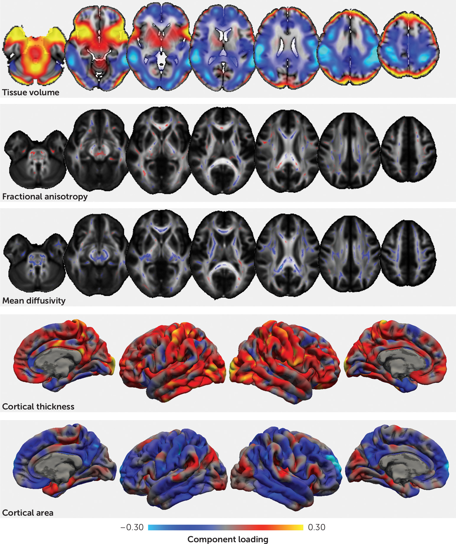
The second independent relationship reveals an association between indices of development and an imaging pattern comprising lower tissue volume in occipital, parietal, and superior frontal regions, greater volume of the anterior temporal lobe, widespread lower mean diffusivity, greater cortical thickness in primary motor and sensory cortical areas and the occipital cortex, and decreased cortical surface area in the frontal, temporal, and parietal cortex (Figure 1). The phenotypic factor was associated primarily with increased body weight (loading=0.88, 95% CI=0.86, 0.93; explained variance, 57%), age (loading=0.31, 95% CI=0.20, 0.43; explained variance, 7%), and pubertal status (loading=0.34, 95% CI=0.24, 0.44; explained variance, 9%). Clinically, the factor is correlated with fewer hyperactive symptoms and a lower probability of receiving medication for ADHD (see Table S3).
FIGURE 1. Spatial Maps Showing the Voxelwise Correlation Between Each Imaging Modality and the Second Imaging Marker Representing Developmenta
a Each imaging marker is represented by a set of spatial maps, one per imaging modality. The weights represent the voxel/vertex-wise correlation between the original imaging data and the respective canonical variates for each multivariate association. Voxelwise correlations between the second canonical imaging variate (canonical pair 2) and each imaging modality are shown. The strength and direction of the correlation is indicated by color. The corresponding phenotypic factor structure is presented in Table S3 in the online supplement. Unthresholded interactive maps are available to view and download on NeuroVault (https://neurovault.org/collections/2277/).

ADHD symptoms.

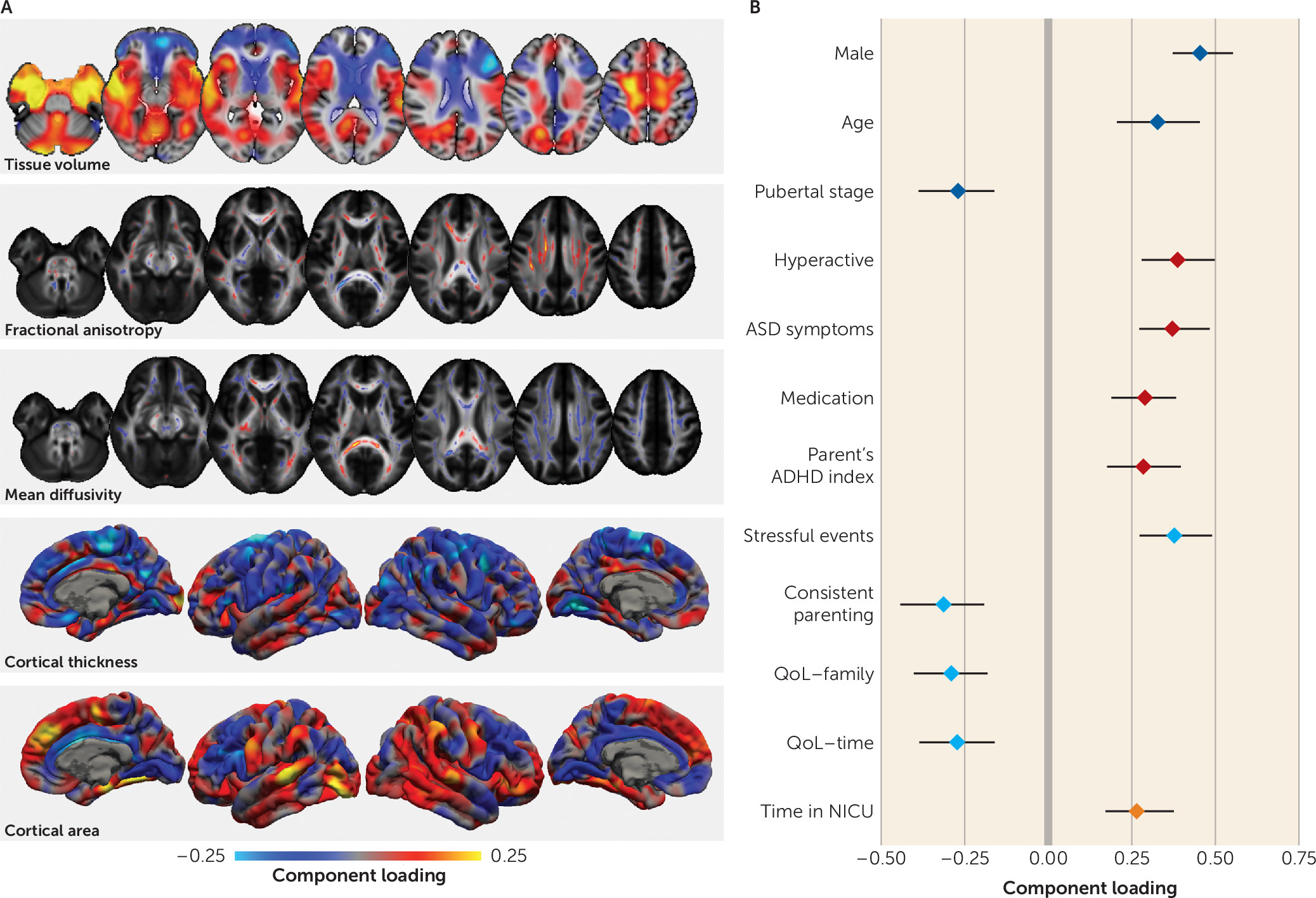
The third relationship describes an imaging pattern associated with ADHD symptoms (Figure 2; see also Table S3). The pattern comprises higher volumes in premotor and temporal regions and the cerebellum and lower volumes in dorsolateral frontal regions, the caudate, and the thalamus. Surface area is lower along the cingulum and prefrontal cortex but higher in the medial superior temporal cortex and dorsomedial frontal cortex. Cortical thickness is lower bilaterally in premotor regions and the anterior cingulate. In white matter, FA is higher in the fornix and the superior longitudinal fasciculus and lower in the genu of the corpus callosum. The associated phenotypic factor is positively correlated with being older (age loading=0.33, 95% CI=0.21, 0.45) but less pubertally developed (loading=−0.27, 95% CI=−0.39, −0.16) and being male (loading=0.45, 95% CI=0.37, 0.55) (see Figure 2B; see also Table S3 in the online supplement). Clinically, it is associated with increased hyperactivity (loading=0.39, 95% CI=0.28, 0.50), with the correlated imaging pattern explaining approximately 8% of variance in hyperactivity symptom count across the cohort (see Table S3), and, to a lesser extent, with increased inattentiveness (loading=0.19, 95% CI=0.08, 0.30) as well as both teacher- and parent-rated ADHD symptom severity indices and an increased probability of receiving ADHD medication. Regarding other clinical features, this factor is associated with more autism spectrum symptoms and social problems, more externalizing but fewer internalizing symptoms, and higher irritability. The associated cognitive profile reflects lower IQ and poorer mathematics and visuospatial reasoning scores. Lower parental education, less consistent parenting style, and lower quality of life were all associated with this factor (see Table S3).
FIGURE 2. Spatial Maps Showing Voxelwise Correlations Between Each Imaging Modality and the Third Imaging Marker and Associated Phenotypic Factor Loadings for the Relationship With ADHD Symptoms and Associated Risk Factorsa
a Voxelwise correlations (color bar) between the third canonical imaging variate (canonical pair 3) and each imaging modality are shown in panel A. Corresponding phenotypic loadings are shown in panel B for individual (blue), clinical (red), familial (cyan), and perinatal (orange) factors (bars indicate 95% confidence interval). Only factors that passed correction for multiple comparisons are shown (p<0.001). The full canonical structure is presented in Table S3 in the online supplement. Unthresholded interactive maps are available to view and download on NeuroVault (https://neurovault.org/collections/2277/). ASD=autistic spectrum disorder; NICU=neonatal intensive care unit; parent’s ADHD index=parent-rated Conners 3 ADHD Index of the child’s behaviors; QoL–family=quality of life, how often child’s behavior interrupted various everyday family activities; QoL–time=quality of life, how often child’s behavior caused cancellation or change of plans (personal or work) at last minute.

Cognitive performance.

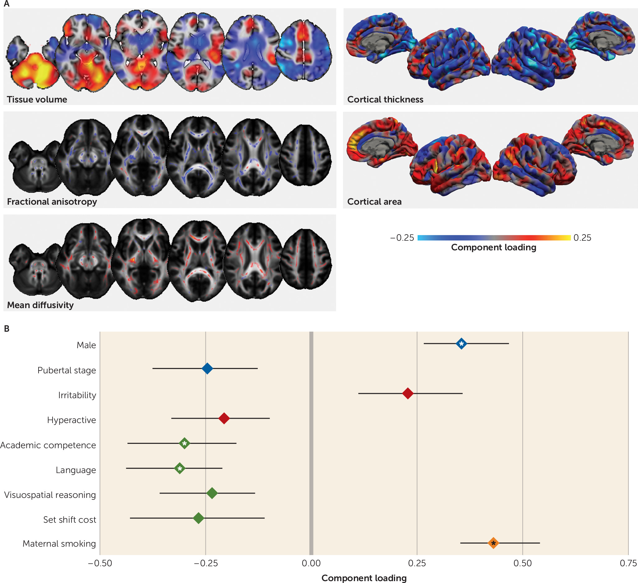
The fourth imaging pattern comprises increased volume in the anterior cingulate and cerebellum, lower volume in the superior lateral frontal and parietal lobes, lower ventricular volume, widespread lower FA and higher MD, lower thickness in lateral aspects of the sensory cortex, and greater cortical area in the medial frontal cortex (Figure 3). The associated phenotypic factor is associated with poorer cognitive performance, marked by lower language scores (loading=−0.31, 95% CI=−0.44, −0.21; explained variance, 4%), reading ability (loading=−0.18, 95% CI=−0.31, −0.05), visuospatial reasoning (loading=−0.23, 95% CI=−0.36, −0.13), and academic competence (loading=−0.30, 95% CI=−0.44, −0.18) but not overall IQ. This factor was also positively associated with male sex, lower parental education, and increased irritability but negatively associated with hyperactive symptoms. There was a strong association with maternal smoking during pregnancy (loading=0.43, 95% CI=0.35, 0.54).
FIGURE 3. Spatial Maps Showing Voxelwise Correlations Between Each Imaging Modality and the Fourth Imaging Marker and Associated Phenotypic Factor Loadings for the Relationship With Cognitive Performancea
a Voxelwise correlations between the fourth canonical imaging variate and each imaging modality are shown in panel A. Phenotypic loadings are shown in panel B for individual (blue), clinical (red), cognitive (green), and perinatal (orange) factors (bars indicate 95% confidence interval). Factor loadings are significant at p<0.01, and factors that passed correction for multiple comparisons are highlighted with an asterisk. The full canonical structure is presented in Table S3 in the online supplement. Unthresholded interactive maps are available to view and download on NeuroVault (https://neurovault.org/collections/2277/).

Model stability and motion considerations.

Overall, the factor structure of the model was robust to different preprocessing strategies, model parameters, and the inclusion of head motion measures (see the Supplemental Results section in the online supplement).

Validation Data Set

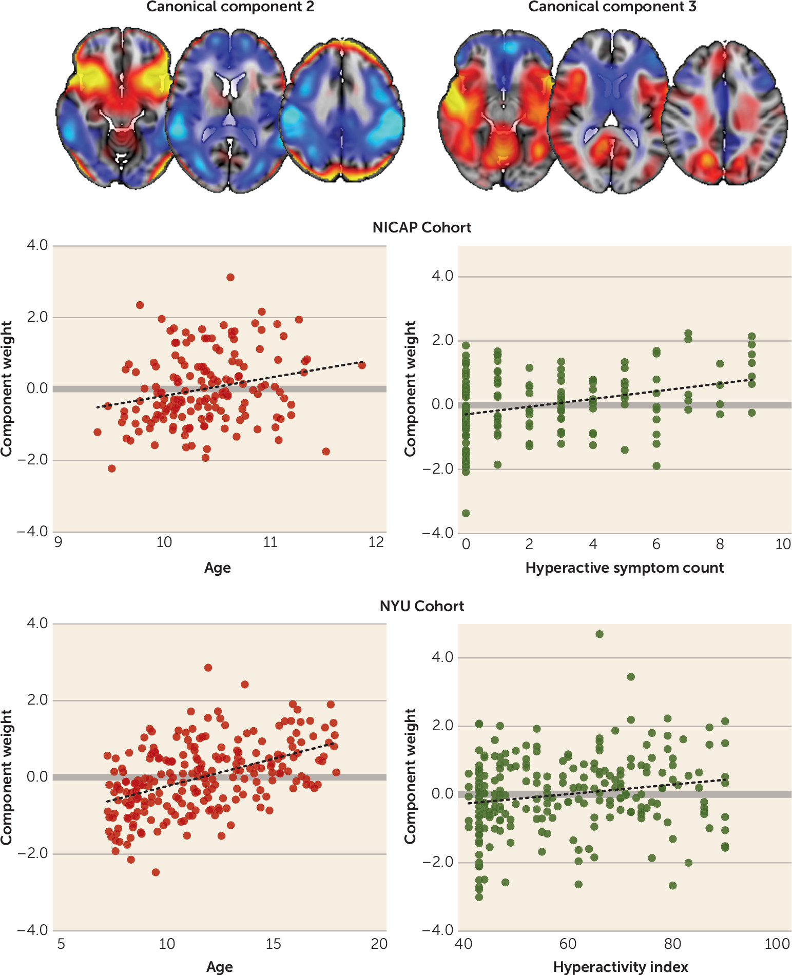
Using imaging patterns derived from the NICAP cohort, we regressed the spatial loading maps onto tissue volume data from the NYU cohort to estimate a set of weights (one per subject) representing the typicality of each pattern for that subject’s imaging data (Figure 4). We found significant linear relationships between the weight of the first three imaging patterns and corresponding phenotypic data in the validation cohort.
FIGURE 4. Validation of Imaging Markers Associated With Age and Hyperactivity, Derived From the Canonical Correlation Analysis, in an Independent Cohorta
a Tissue volume loading maps are shown for canonical pairs 2 and 3 (top row). In the Children’s Attention Project neuroimaging (NICAP) cohort, component weights were associated with age and hyperactive symptom count, respectively (middle row). Using multivariate regression, expression of each imaging pattern was estimated in an independent data set. Imaging marker expression was found to predict age and hyperactivity index in the New York University (NYU) cohort (bottom row).
As expected, the imaging pattern reflecting head size was strongly correlated with intracranial volume in the NYU cohort (R2=0.86; F=1460.0, df=1, 229, p<0.001). We also found a significant linear relationship between the second imaging pattern (reflecting development) (Figure 1) and age (R2=0.23; F=67.96, df=1, 229, p<0.001) in the validation cohort. This marker did not correlate with ADHD index (p=0.31), hyperactivity (p=0.14), inattentiveness (p=0.69), or IQ (p=0.63).
In contrast, the third imaging pattern (reflecting ADHD symptoms) significantly predicted hyperactivity (R2=0.04, F=9.12, p=0.003) (Figure 4) and, to a lesser extent, inattention (R2=0.02, F=5.49, p=0.02) in the NYU cohort, but it did not predict age (p=0.14) or IQ (p=0.31). The inclusion of sex, age, and IQ in the model with marker strength explained almost 10% of variance in hyperactivity score in the NYU cohort (R2=0.097; see the Supplemental Results section in the online supplement). Secondary to the main aim of this study, we also found that the expression of this imaging pattern was significantly different between ADHD diagnostic groups in both the NICAP and NYU cohorts.

Discussion

In a data-driven analysis of multimodal imaging data and multi-informant phenotypic data, we identified a novel set of brain imaging patterns that reflect phenotypic variation across a cohort of children with ADHD and non-ADHD control subjects. Our results highlight how multiple cognitive and clinical variables can be factored to explain observed behaviors. Importantly, the inclusion of neuroimaging data allows the identification and separation of the neurobiological correlates of these factors. Patterns across multiple anatomical scales were associated with developmental, clinical, and cognitive profiles. We found that these patterns proved informative to models of phenotypic variation in an independent pediatric cohort.
The first imaging pattern captured the well-known relationship between brain volume and cortical surface area (39). Intracranial volume was higher in males, and individuals with higher intracranial volume scored well in mathematics and reading, lending support to recent reports of the shared genetic etiology between brain size, cognitive ability, and educational attainment (40). This pattern was positively correlated with socioeconomic status, mirroring previous reports (41). Contrary to evidence that intracranial volume is reduced in ADHD (9, 18), ADHD symptoms were not significantly associated with this imaging marker. However, given the relatively small effect size of this reported difference, this is perhaps not surprising (18).
The second pattern captured neuroanatomical variation associated with childhood development indexed by greater body weight, pubertal status, and age. During puberty, longitudinal studies report decreases in cortical thickness with increasing circulating testosterone (42). However, in prepubertal cohorts, dehydroepiandrosterone (DHEA), rather than testosterone, is associated with brain growth. DHEA levels have been associated with increased cortical thickness in the dorsolateral prefrontal cortex, the premotor cortex, and the temporoparietal junction in both males and females (43). We previously showed that pubertal onset is associated with increased fiber density in the corpus callosum (44). In the present study, we found a pattern of lower mean diffusivity evident in both the corpus callosum and the ascending white matter tracts. We postulate that the changes reported here reflect a combination of pubertal processes in addition to age-related variation.
We also observed a significant association between the second phenotypic factor and medication status, suggesting that younger, less pubertally mature children were more likely to be receiving medication for ADHD. While this is broadly consistent with epidemiological evidence showing that younger age relative to peers increases the likelihood of ADHD diagnosis and subsequent medication (28), we previously reported that there is little evidence to support a simple correlation between age and diagnosis in this cohort (45). Indeed, Table S3 in the online supplement shows that although a lower score for this factor is associated with a slight increase in hyperactive symptoms (loading=−0.17), it explains only a small amount of variance in hyperactive symptom count (approximately 2%) and it occurs independently of other clinical factors. We propose that this pattern instead represents a marker of lower neuroanatomical maturity—or “brain age” (46)—that is associated with both age and pubertal status and may manifest as a more immature behavioral profile characterized partly by increased hyperactivity.
The third imaging pattern was correlated with a clinical profile marked by higher hyperactive symptom count, higher teacher and parent ratings of ADHD behavior, and more comorbidities. This profile was more common among male participants, who were less pubertally developed. It also correlated less with inattentive symptoms than with hyperactive symptoms, suggesting some domain specificity.
Previous studies comparing individuals with ADHD with control subjects have reported similar neuroimaging differences in clinical populations, including volumetric decreases in the anterior cingulate cortex and increases in the temporal lobe and around sensorimotor regions that covaried with hyperactive symptom severity (23, 24). Other univariate approaches have yielded differences in frontal, parietal, and subcortical structures consistent with the patterns described here (9, 10, 47). Our results also highlight the role of the superior longitudinal fasciculus, a structure that has been implicated in ADHD (48), particularly in relation to hyperactivity (49).
By including a rich scope of phenotypic data, we found that this factor was also correlated with poorer academic performance, lower parental education, and an increased experience of stressful life events. Symptoms of inattention and hyperactivity have previously been shown to correlate inversely with academic performance (27), and early life deprivation can result in neuroanatomical alterations that mirror those seen in ADHD (50). Similarly, van der Meer et al. (51) identified genetic variants associated with stress exposure and hypothalamic-pituitary-adrenal axis activity that predicted ADHD severity in childhood, explaining approximately 12.5% of variance in symptom severity. These findings highlight possible mechanistic pathways that may lead to altered developmental neuroanatomy in ADHD.
To demonstrate the external validity of our model, we estimated the expression of each imaging pattern in an independent cohort. We found a small but significant independent association between the ADHD imaging pattern and hyperactivity index in this validation cohort. In combination with age, sex, and IQ, this model explained approximately 10% of the variance in hyperactivity score in the NYU cohort. The imaging pattern alone explained 8% of variance in hyperactivity in the NICAP cohort. We also observed a measure of symptom specificity, with the imaging marker explaining approximately twice as much variation in hyperactivity than in inattentiveness in both the NICAP and NYU cohorts. The model compares well to previous reports. Recent large-scale genetic analyses have demonstrated that markers of genetic risk for ADHD explain around 0.1%–0.5% of the variance in ADHD trait behavior in the general population (52). Furthermore, longitudinal studies find that neuropsychological assessment in preschool, socioeconomic status, and sex together explain around 8%–12% of variance in ADHD symptom severity in the same individual in later childhood (53). With regard to MRI, Rosenberg et al. (54) derived an imaging marker from functional MRI (fMRI) scans acquired during an attention task and found that this marker explained approximately 7%–9% of variance in ADHD symptom severity in an independent population.
The fourth imaging pattern was associated with a poorer cognitive profile but not IQ, and it highlighted the role of the dorsal attention networks and associated white matter organization for performance in visuospatial tasks (55, 56). Although there were no broader cognitive variables available to test in the validation cohort, IQ was also not associated with expression of this marker in the NYU cohort. This suggests that this factor is not reflective of a general decrease in cognitive functioning but rather is specific to certain domains. Although some aspects of this factor structure proved less stable under model perturbation than the preceding three (see Figure S4 in the online supplement), an environmental component may underlie cognitive performance, with lower parental education, maternal smoking, and a poorer quality of life associated with the cognitive profile. Clinically, the factor was associated with increased irritability and fewer hyperactive symptoms. Irritability is a common comorbid factor in ADHD, although this analysis suggests that its underlying neural correlates may be independent of ADHD, supporting a recent meta-analysis that suggests that irritability is a poor predictor of ADHD (57).
In summary, these findings suggest that the presentation of ADHD may arise from a summation of several clinical, developmental, or cognitive factors, each with a distinct neuroanatomical foundation. Separate from the main clinical relationship, one factor reflected delayed development relative to peers, consistent with the theory of delayed maturation in ADHD (29). Similarly, although irritability is often present in ADHD, it also had a presentation, independent of ADHD, associated with its own pattern of cognitive deficits and anatomical correlates. We anticipate that our work will inform future research into the neurobiological foundations of ADHD, highlighting the value of neuroimaging and the importance of leveraging detailed phenotypic data to understand the neurobiology underlying neurodevelopmental disorders.
Our study has a number of limitations that warrant consideration. The age range of the participants was relatively narrow, and the reported brain-behavior associations may vary across different stages of development. Future research in this longitudinal cohort will examine whether changes in these imaging markers predict longitudinal change in developmental, clinical, and cognitive phenotypes. Twenty-three individuals were taking medication for their ADHD, and the long-term effect of medication on brain structure may constitute a confounding factor. However, as medication may normalize neuroanatomical differences in ADHD (58), such an effect would be expected to reduce the magnitude of the findings.
We plan in future work to incorporate functional brain measures derived from fMRI into this analytical framework. Co-occurring functional-structural deficits have been reported in ADHD using multimodal methods (25), and fMRI networks are able to predict variation in attention and classify DHD subtypes (54, 59). We anticipate that characterizing the functional correlates of the neuroanatomical patterns described here will prove informative to the theoretical modeling of the disorder.
In conclusion, this study furthers our understanding of brain development and mental health in childhood with evidence derived from multidimensional phenotypic and neurobiological measures. We identified four separable neuroanatomical patterns that are associated with unique developmental, clinical, and cognitive factors that span diagnostic criteria. We further demonstrated that the markers associated with development and an ADHD phenotype derived from a community sample can predict phenotypic variation in an independent cohort.
Acknowledgments

Dr. Sciberras is supported by an NHMRC Career Development Fellowship (1110688). Dr. Anderson was supported by an NHMRC Senior Practitioner Fellowship (1079478). Dr. Nicholson was funded by the Australian Communities Foundation through the Roberta Holmes Chair for the Transition to Contemporary Parenthood Program (Coronella subfund). Dr. Efron is supported by a Clinical Scientist Fellowship from the Murdoch Children’s Research Institute.
The authors thank the Royal Children’s Hospital medical imaging staff for their assistance and expertise in the collection of the MRI data included in this study, and the many families and schools for their participation in the study.

Footnote

This study was funded by project grant 1065895 from the National Health and Medical Research Council of Australia (NHMRC). The Children’s Attention Project was funded by NHMRC project grant 1008522 and a grant from the Collier Foundation. The research was conducted within the Developmental Imaging research group, Murdoch Children’s Research Institute and the Children’s MRI Centre, Royal Children’s Hospital, Melbourne. It was supported by the Murdoch Children’s Research Institute, the Royal Children’s Hospital, the Royal Children’s Hospital Foundation, the Department of Paediatrics at the University of Melbourne, and the Victorian Government’s Operational Infrastructure Support Program. The authors also thank Dr. Michael P. Milham and the ADHD-200 consortium for making the ADHD-200 data set available to researchers. Funding for the NYU cohort was provided by NIMH (R01MH083246), Autism Speaks, the Stavros Niarchos Foundation, the Leon Levy Foundation, and an endowment provided by Phyllis Green and Randolph Cōwen.

Supplementary Material

File (appi.ajp.2018.18010034.ds001.pdf)

References

1.
Insel TR: The NIMH Research Domain Criteria (RDoC) Project: precision medicine for psychiatry. Am J Psychiatry 2014; 171:395–397
2.
Woo C-W, Chang LJ, Lindquist MA, et al: Building better biomarkers: brain models in translational neuroimaging. Nat Neurosci 2017; 20:365–377
3.
Polanczyk G, de Lima MS, Horta BL, et al: The worldwide prevalence of ADHD: a systematic review and metaregression analysis. Am J Psychiatry 2007; 164:942–948
4.
Barkley RA: Behavioral inhibition, sustained attention, and executive functions: constructing a unifying theory of ADHD. Psychol Bull 1997; 121:65–94
5.
Berquin PC, Giedd JN, Jacobsen LK, et al: Cerebellum in attention-deficit hyperactivity disorder: a morphometric MRI study. Neurology 1998; 50:1087–1093
6.
Silk TJ, Vance A, Rinehart N, et al: White-matter abnormalities in attention deficit hyperactivity disorder: a diffusion tensor imaging study. Hum Brain Mapp 2009; 30:2757–2765
7.
Frodl T, Skokauskas N: Meta-analysis of structural MRI studies in children and adults with attention deficit hyperactivity disorder indicates treatment effects. Acta Psychiatr Scand 2012; 125:114–126
8.
Valera EM, Faraone SV, Murray KE, et al: Meta-analysis of structural imaging findings in attention-deficit/hyperactivity disorder. Biol Psychiatry 2007; 61:1361–1369
9.
Silk TJ, Beare R, Malpas C, et al: Cortical morphometry in attention deficit/hyperactivity disorder: contribution of thickness and surface area to volume. Cortex 2016; 82:1–10
10.
Shaw P, Malek M, Watson B, et al: Development of cortical surface area and gyrification in attention-deficit/hyperactivity disorder. Biol Psychiatry 2012; 72:191–197
11.
Wolosin SM, Richardson ME, Hennessey JG, et al: Abnormal cerebral cortex structure in children with ADHD. Hum Brain Mapp 2009; 30:175–184
12.
Shaw P, Malek M, Watson B, et al: Trajectories of cerebral cortical development in childhood and adolescence and adult attention-deficit/hyperactivity disorder. Biol Psychiatry 2013; 74:599–606
13.
van Ewijk H, Heslenfeld DJ, Zwiers MP, et al: Diffusion tensor imaging in attention deficit/hyperactivity disorder: a systematic review and meta-analysis. Neurosci Biobehav Rev 2012; 36:1093–1106
14.
Cooper M, Thapar A, Jones DK: ADHD severity is associated with white matter microstructure in the subgenual cingulum. Neuroimage Clin 2015; 7:653–660
15.
Cortese S, Kelly C, Chabernaud C, et al: Toward systems neuroscience of ADHD: a meta-analysis of 55 fMRI studies. Am J Psychiatry 2012; 169:1038–1055
16.
Konrad K, Eickhoff SB: Is the ADHD brain wired differently? A review on structural and functional connectivity in attention deficit hyperactivity disorder. Hum Brain Mapp 2010; 31:904–916
17.
Uddin LQ, Dajani DR, Voorhies W, et al: Progress and roadblocks in the search for brain-based biomarkers of autism and attention-deficit/hyperactivity disorder. Transl Psychiatry 2017; 7:e1218
18.
Hoogman M, Bralten J, Hibar DP, et al: Subcortical brain volume differences in participants with attention deficit hyperactivity disorder in children and adults: a cross-sectional mega-analysis. Lancet Psychiatry 2017; 4:310–319
19.
Tamnes CK, Ostby Y, Fjell AM, et al: Brain maturation in adolescence and young adulthood: regional age-related changes in cortical thickness and white matter volume and microstructure. Cereb Cortex 2010; 20:534–548
20.
Wu M, Lu LH, Lowes A, et al: Development of superficial white matter and its structural interplay with cortical gray matter in children and adolescents. Hum Brain Mapp 2014; 35:2806–2816
21.
Van Essen DC: A tension-based theory of morphogenesis and compact wiring in the central nervous system. Nature 1997; 385:313–318
22.
Groves AR, Beckmann CF, Smith SM, et al: Linked independent component analysis for multimodal data fusion. Neuroimage 2011; 54:2198–2217
23.
Francx W, Llera A, Mennes M, et al: Integrated analysis of gray and white matter alterations in attention-deficit/hyperactivity disorder. Neuroimage Clin 2016; 11:357–367
24.
Wolfers T, Arenas AL, Onnink AMH, et al: Refinement by integration: aggregated effects of multimodal imaging markers on adult ADHD. J Psychiatry Neurosci 2017; 42:386–394
25.
Kessler D, Angstadt M, Welsh RC, et al: Modality-spanning deficits in attention-deficit/hyperactivity disorder in functional networks, gray matter, and white matter. J Neurosci 2014; 34:16555–16566
26.
Spencer TJ, Biederman J, Mick E: Attention-deficit/hyperactivity disorder: diagnosis, lifespan, comorbidities, and neurobiology. J Pediatr Psychol 2007; 32:631–642
27.
Merrell C, Tymms PB: Inattention, hyperactivity, and impulsiveness: their impact on academic achievement and progress. Br J Educ Psychol 2001; 71:43–56
28.
Whitely M, Lester L, Phillimore J, et al: Influence of birth month on the probability of Western Australian children being treated for ADHD. Med J Aust 2017; 206:85
29.
El-Sayed E, Larsson JO, Persson HE, et al: “Maturational lag” hypothesis of attention deficit hyperactivity disorder: an update. Acta Paediatr 2003; 92:776–784
30.
Silk TJ, Genc S, Anderson V, et al: Developmental brain trajectories in children with ADHD and controls: a longitudinal neuroimaging study. BMC Psychiatry 2016; 16:59
31.
Sciberras E, Efron D, Schilpzand EJ, et al: The Children’s Attention Project: a community-based longitudinal study of children with ADHD and non-ADHD controls. BMC Psychiatry 2013; 13:18
32.
Rueckert D, Frangi AF, Schnabel JA: Automatic construction of 3-D statistical deformation models of the brain using nonrigid registration. IEEE Trans Med Imaging 2003; 22:1014–1025
33.
Fischl B: FreeSurfer. Neuroimage 2012; 62:774–781
34.
Smith SM, Jenkinson M, Woolrich MW, et al: Advances in functional and structural MR image analysis and implementation as FSL. Neuroimage 2004; 23(suppl 1):S208–S219
35.
Smith SM, Jenkinson M, Johansen-Berg H, et al: Tract-based spatial statistics: voxelwise analysis of multi-subject diffusion data. Neuroimage 2006; 31:1487–1505
36.
Ball G, Aljabar P, Nongena P, et al: Multimodal image analysis of clinical influences on preterm brain development. Ann Neurol 2017; 82:233–246
37.
Bartlett MS: The statistical significance of canonical correlations. Biometrika 1941; 32:29–37
38.
ADHD-200 Consortium: A model to advance the translational potential of neuroimaging in clinical neuroscience. Front Syst Neurosci (eCollection, Sep 5, 2012). https://www.ncbi.nlm.nih.gov/pmc/articles/PMC3433679/
39.
Toro R, Perron M, Pike B, et al: Brain size and folding of the human cerebral cortex. Cereb Cortex 2008; 18:2352–2357
40.
Hagenaars SP, Harris SE, Davies G, et al: Shared genetic aetiology between cognitive functions and physical and mental health in UK Biobank (N=112 151) and 24 GWAS consortia. Mol Psychiatry 2016; 21:1624–1632
41.
Noble KG, Houston SM, Brito NH, et al: Family income, parental education, and brain structure in children and adolescents. Nat Neurosci 2015; 18:773–778
42.
Nguyen T-V, McCracken J, Ducharme S, et al: Testosterone-related cortical maturation across childhood and adolescence. Cereb Cortex 2013; 23:1424–1432
43.
Nguyen T-V, McCracken JT, Ducharme S, et al: Interactive effects of dehydroepiandrosterone and testosterone on cortical thickness during early brain development. J Neurosci 2013; 33:10840–10848
44.
Genc S, Seal ML, Dhollander T, et al: White matter alterations at pubertal onset. Neuroimage 2017; 156:286–292
45.
Sciberras E, Gulenc A, Efron D: Influence of birth month on the probability of Western Australian children being treated for ADHD. Med J Aust 2017; 207:268
46.
Erus G, Battapady H, Satterthwaite TD, et al: Imaging patterns of brain development and their relationship to cognition. Cereb Cortex 2015; 25:1676–1684
47.
Batty MJ, Liddle EB, Pitiot A, et al: Cortical gray matter in attention-deficit/hyperactivity disorder: a structural magnetic resonance imaging study. J Am Acad Child Adolesc Psychiatry 2010; 49:229–238
48.
Makris N, Buka SL, Biederman J, et al: Attention and executive systems abnormalities in adults with childhood ADHD: A DT-MRI study of connections. Cereb Cortex 2008; 18:1210–1220
49.
Mostofsky SH, Simmonds DJ: Response inhibition and response selection: two sides of the same coin. J Cogn Neurosci 2008; 20:751–761
50.
McLaughlin KA, Sheridan MA, Winter W, et al: Widespread reductions in cortical thickness following severe early-life deprivation: a neurodevelopmental pathway to attention-deficit/hyperactivity disorder. Biol Psychiatry 2014; 76:629–638
51.
van der Meer D, Hoekstra PJ, van Donkelaar M, et al: Predicting attention-deficit/hyperactivity disorder severity from psychosocial stress and stress-response genes: a random forest regression approach. Transl Psychiatry 2017; 7:e1145
52.
Martin J, Hamshere ML, Stergiakouli E, et al: Genetic risk for attention-deficit/hyperactivity disorder contributes to neurodevelopmental traits in the general population. Biol Psychiatry 2014; 76:664–671
53.
Sjöwall D, Bohlin G, Rydell A-M, et al: Neuropsychological deficits in preschool as predictors of ADHD symptoms and academic achievement in late adolescence. Child Neuropsychol 2017; 23:111–128
54.
Rosenberg MD, Finn ES, Scheinost D, et al: A neuromarker of sustained attention from whole-brain functional connectivity. Nat Neurosci 2016; 19:165–171
55.
Umarova RM, Saur D, Schnell S, et al: Structural connectivity for visuospatial attention: significance of ventral pathways. Cereb Cortex 2010; 20:121–129
56.
Bressler SL, Tang W, Sylvester CM, et al: Top-down control of human visual cortex by frontal and parietal cortex in anticipatory visual spatial attention. J Neurosci 2008; 28:10056–10061
57.
Vidal-Ribas P, Brotman MA, Valdivieso I, et al: The status of irritability in psychiatry: a conceptual and quantitative review. J Am Acad Child Adolesc Psychiatry 2016; 55:556–570
58.
Spencer TJ, Brown A, Seidman LJ, et al: Effect of psychostimulants on brain structure and function in ADHD: a qualitative literature review of magnetic resonance imaging-based neuroimaging studies. J Clin Psychiatry 2013; 74:902–917
59.
Fair DA, Nigg JT, Iyer S, et al: Distinct neural signatures detected for ADHD subtypes after controlling for micro-movements in resting state functional connectivity MRI data. Front Syst Neurosci 2013; 6:80

Information & Authors

Information

Published In

Go to American Journal of Psychiatry
American Journal of Psychiatry
Pages: 57 - 66
PubMed: 30220220

History

Received: 10 January 2018
Revision received: 24 April 2018
Revision received: 25 May 2018
Revision received: 1 June 2018
Accepted: 11 June 2018
Published online: 17 September 2018
Published in print: January 01, 2019

Keywords

  1. Attention Deficit Hyperactivity Disorder
  2. Brain Imaging Techniques

Authors

Details

Gareth Ball, Ph.D. [email protected]
From Developmental Imaging, Clinical Sciences, Murdoch Children’s Research Institute, Melbourne, Australia; Population Health, Murdoch Children’s Research Institute, Melbourne; the Royal Children’s Hospital, Melbourne; the Department of Paediatrics, University of Melbourne, Melbourne; the Judith Lumley Centre, La Trobe University, Melbourne; the School of Psychology, Deakin University, Geelong, Australia; and the Melbourne School of Psychological Sciences, University of Melbourne, Melbourne.
Charles B. Malpas, Ph.D.
From Developmental Imaging, Clinical Sciences, Murdoch Children’s Research Institute, Melbourne, Australia; Population Health, Murdoch Children’s Research Institute, Melbourne; the Royal Children’s Hospital, Melbourne; the Department of Paediatrics, University of Melbourne, Melbourne; the Judith Lumley Centre, La Trobe University, Melbourne; the School of Psychology, Deakin University, Geelong, Australia; and the Melbourne School of Psychological Sciences, University of Melbourne, Melbourne.
Sila Genc, M.Sc.
From Developmental Imaging, Clinical Sciences, Murdoch Children’s Research Institute, Melbourne, Australia; Population Health, Murdoch Children’s Research Institute, Melbourne; the Royal Children’s Hospital, Melbourne; the Department of Paediatrics, University of Melbourne, Melbourne; the Judith Lumley Centre, La Trobe University, Melbourne; the School of Psychology, Deakin University, Geelong, Australia; and the Melbourne School of Psychological Sciences, University of Melbourne, Melbourne.
Daryl Efron, M.D., F.R.A.C.P.
From Developmental Imaging, Clinical Sciences, Murdoch Children’s Research Institute, Melbourne, Australia; Population Health, Murdoch Children’s Research Institute, Melbourne; the Royal Children’s Hospital, Melbourne; the Department of Paediatrics, University of Melbourne, Melbourne; the Judith Lumley Centre, La Trobe University, Melbourne; the School of Psychology, Deakin University, Geelong, Australia; and the Melbourne School of Psychological Sciences, University of Melbourne, Melbourne.
Emma Sciberras, D.Psych.
From Developmental Imaging, Clinical Sciences, Murdoch Children’s Research Institute, Melbourne, Australia; Population Health, Murdoch Children’s Research Institute, Melbourne; the Royal Children’s Hospital, Melbourne; the Department of Paediatrics, University of Melbourne, Melbourne; the Judith Lumley Centre, La Trobe University, Melbourne; the School of Psychology, Deakin University, Geelong, Australia; and the Melbourne School of Psychological Sciences, University of Melbourne, Melbourne.
Vicki Anderson, Ph.D.
From Developmental Imaging, Clinical Sciences, Murdoch Children’s Research Institute, Melbourne, Australia; Population Health, Murdoch Children’s Research Institute, Melbourne; the Royal Children’s Hospital, Melbourne; the Department of Paediatrics, University of Melbourne, Melbourne; the Judith Lumley Centre, La Trobe University, Melbourne; the School of Psychology, Deakin University, Geelong, Australia; and the Melbourne School of Psychological Sciences, University of Melbourne, Melbourne.
Jan M. Nicholson, Ph.D.
From Developmental Imaging, Clinical Sciences, Murdoch Children’s Research Institute, Melbourne, Australia; Population Health, Murdoch Children’s Research Institute, Melbourne; the Royal Children’s Hospital, Melbourne; the Department of Paediatrics, University of Melbourne, Melbourne; the Judith Lumley Centre, La Trobe University, Melbourne; the School of Psychology, Deakin University, Geelong, Australia; and the Melbourne School of Psychological Sciences, University of Melbourne, Melbourne.
[email protected]Timothy J. Silk, Ph.D.
From Developmental Imaging, Clinical Sciences, Murdoch Children’s Research Institute, Melbourne, Australia; Population Health, Murdoch Children’s Research Institute, Melbourne; the Royal Children’s Hospital, Melbourne; the Department of Paediatrics, University of Melbourne, Melbourne; the Judith Lumley Centre, La Trobe University, Melbourne; the School of Psychology, Deakin University, Geelong, Australia; and the Melbourne School of Psychological Sciences, University of Melbourne, Melbourne.

Notes

Address correspondence to Dr. Ball ([email protected]) and Dr. Silk ([email protected]).

Competing Interests

The authors report no financial relationships with commercial interests.

Funding Information

National Health and Medical Research Council10.13039/501100000925: 1008522, 1065895

Metrics & Citations

Metrics

Citations

Export Citations

If you have the appropriate software installed, you can download article citation data to the citation manager of your choice. Simply select your manager software from the list below and click Download.

For more information or tips please see 'Downloading to a citation manager' in the Help menu.

Format
Citation style
Style
Copy to clipboard

View Options

View options

PDF/EPUB

View PDF/EPUB

Login options

Already a subscriber? Access your subscription through your login credentials or your institution for full access to this article.

Personal login Institutional Login Open Athens login
Purchase Options

Purchase this article to access the full text.

PPV Articles - American Journal of Psychiatry

PPV Articles - American Journal of Psychiatry

Not a subscriber?

Subscribe Now / Learn More

PsychiatryOnline subscription options offer access to the DSM-5-TR® library, books, journals, CME, and patient resources. This all-in-one virtual library provides psychiatrists and mental health professionals with key resources for diagnosis, treatment, research, and professional development.

Need more help? PsychiatryOnline Customer Service may be reached by emailing [email protected] or by calling 800-368-5777 (in the U.S.) or 703-907-7322 (outside the U.S.).

Media

Figures

Other

Tables

Share

Share

Share article link

Share